Introduction
This chapter is a photo gallery of igneous and metamorphic rocks. Different parts of the chapter have brief introductions to put the photos in context, but mostly this chapter is meant only to provide many visual examples of many different rocks.
To navigate this chapter, use the links in the table below to get to different sections. For more detailed navigation, use the Table of Contents on the right side of this web page.
What’s Here?
Most rocks in this collection have a hand sample photo, a plane-polars (PP) thin-section view, and a crossed-polars (XP) thin-section view. If you click on an image, you will get an enlarged view which you can close using the back arrow.
Many rocks (and we are adding more) have a link to a 3D rotatable model of the sample. The rotatable images open in a different tab on your browser. To get back to this book, you must close that second tab.
Photo Sources
Many of the photos in this chapter were taken specifically for this book. Others came from existing University of North Dakota websites, and others came from a number of different internet sources. A list of all sources is at the end of this chapter.
16.1 Plutonic Rocks
16.1.1 Quartz-Rich Granitoids
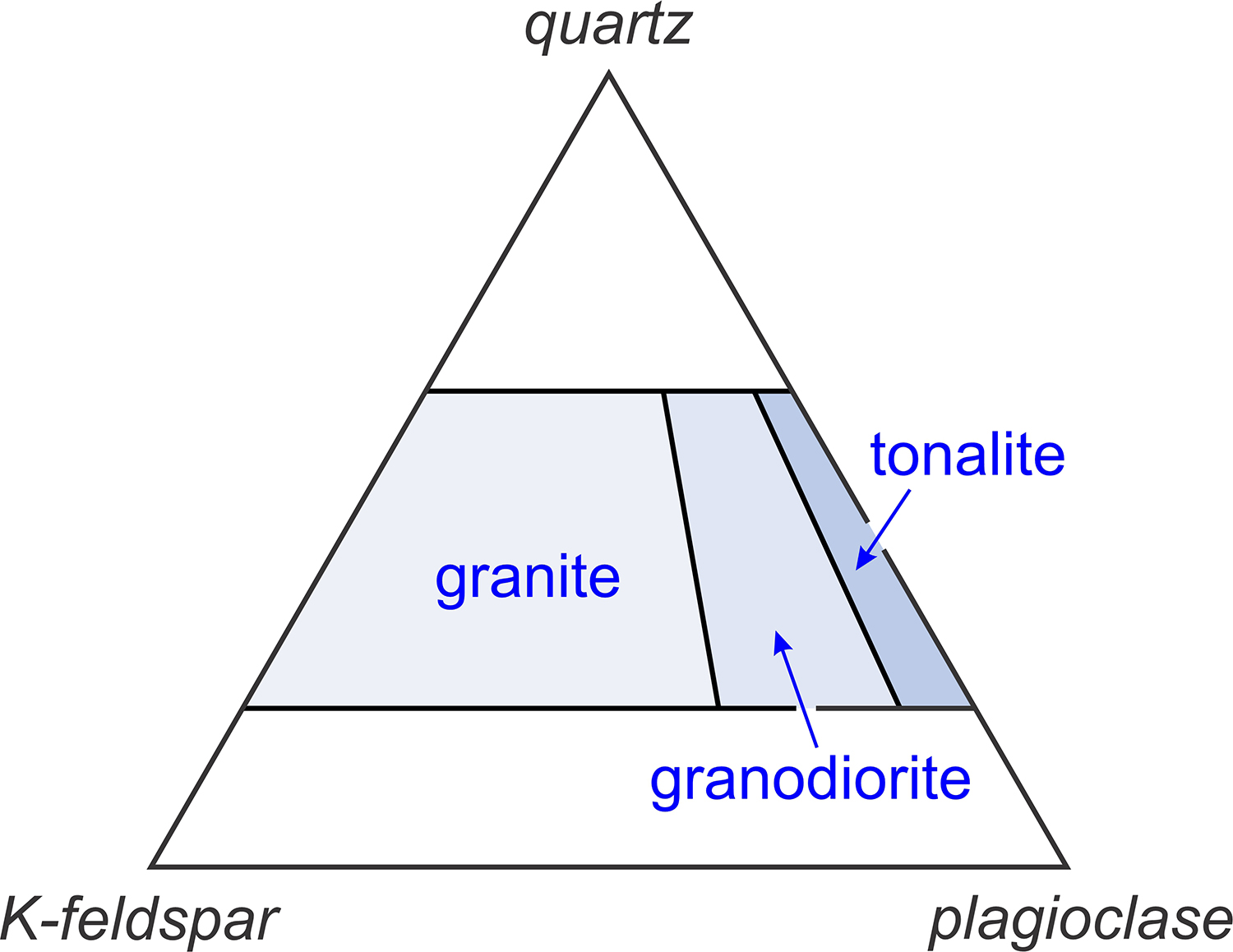
Granitoids are a diverse group of plutonic rocks that contain some combination of essential quartz, plagioclase, and alkali feldspar. Two (or three) of these minerals are always present in granitoids. Quartz-rich granitoids include three main rock types: granites, granodiorites, and tonalites. All contain essential quartz with feldspar. The feldspar can be plagioclase or alkali feldspar, or both, depending on the kind of rock (Figure 16.1). But there are many varieties of these granitoids, varying mostly because they contain different minor minerals. Micas (biotite and sometimes muscovite), and amphiboles or pyroxenes of various sorts, are commonly present. Many other minerals may be present in small amounts.
▪Fine-grained Granite (Aplite)
|
|
|
|
| ⇒Link to 3D rotatable image of this aplite |
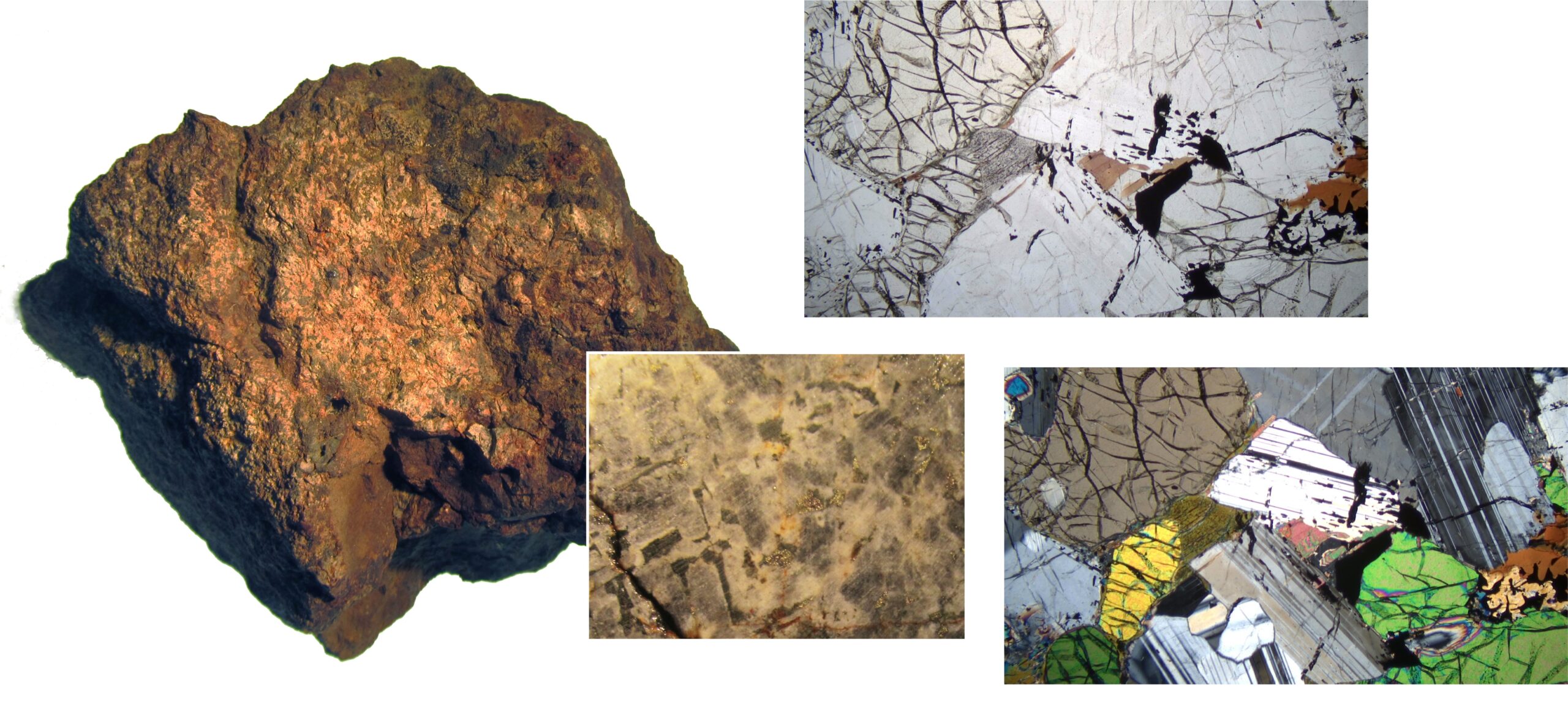
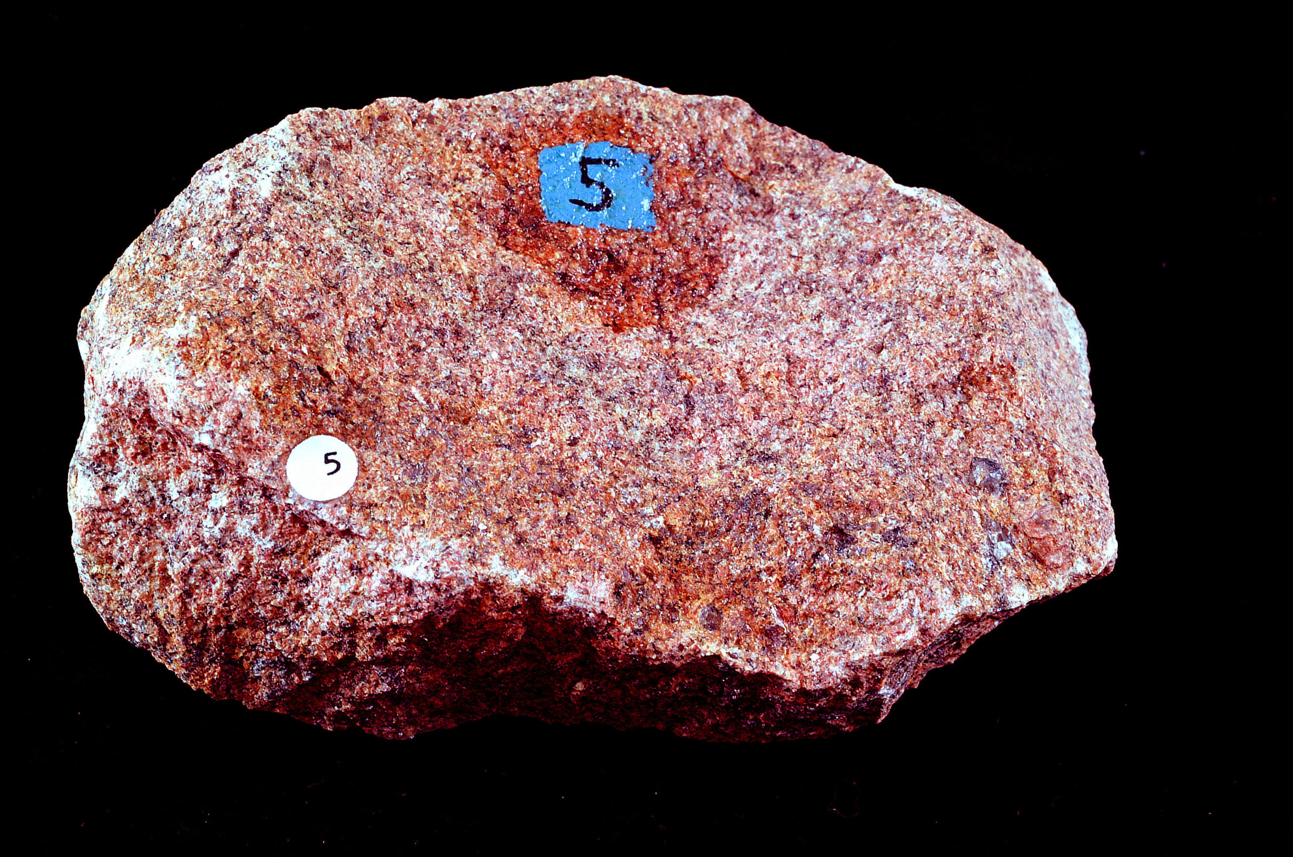
Aplites are rocks equivalent to fine-grained granites. Like all granites, they contain quartz and K-feldspar as essential minerals. The photos above show the Silver Plume aplite from near Boulder, Colorado. The sugary-textured rock contains predominately quartz, microcline, plagioclase, and lesser amounts of biotite. Most crystals in this rock are well under a millimeter in size but two larger quartz crystals – several millimeters across- can be seen near the right corner of the hand sample.
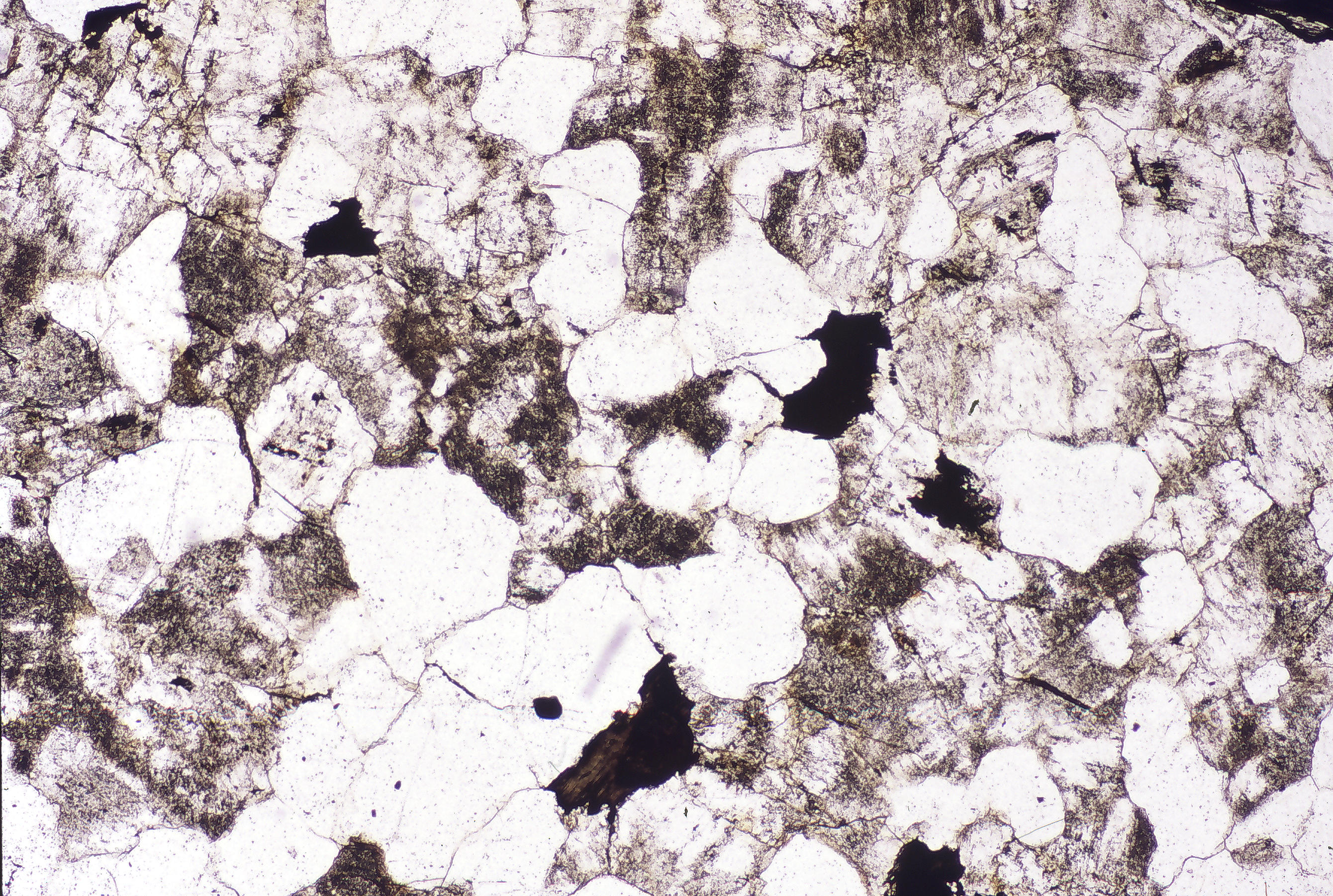
In the plane polars view, the feldspars have rough surface due to incipient alteration; some show traces of twinning. The quartz is clear. Dark colored grains are biotite. In crossed polars, the quartz and feldspar have typical 1st-order interference colors. The biotite has such a strong dark-brown mineral color that interference colors are hard to discern.
▪Biotite-Hornblende Granite
|
|
|
|
| ⇒Link to 3D rotatable image of this granite |
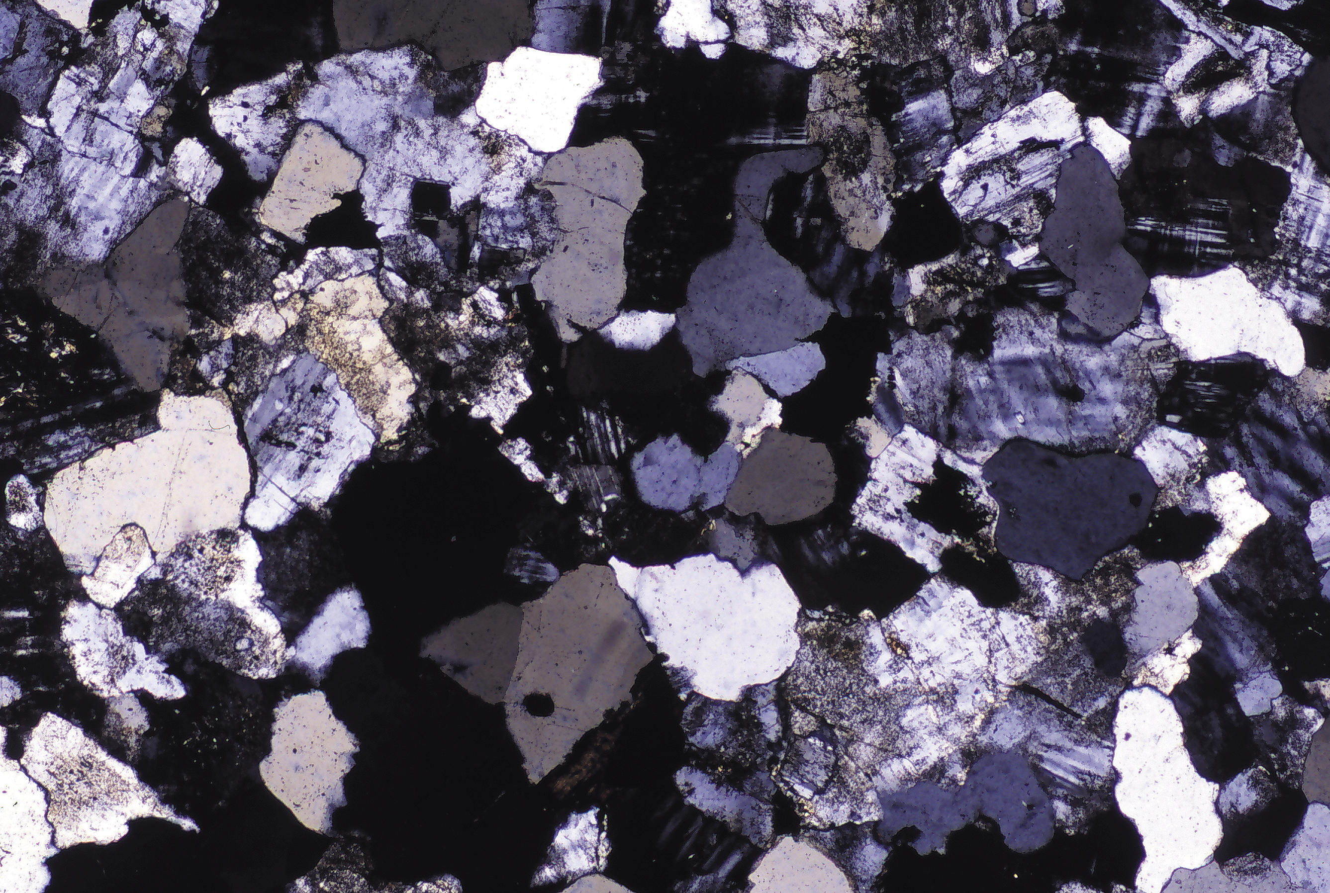
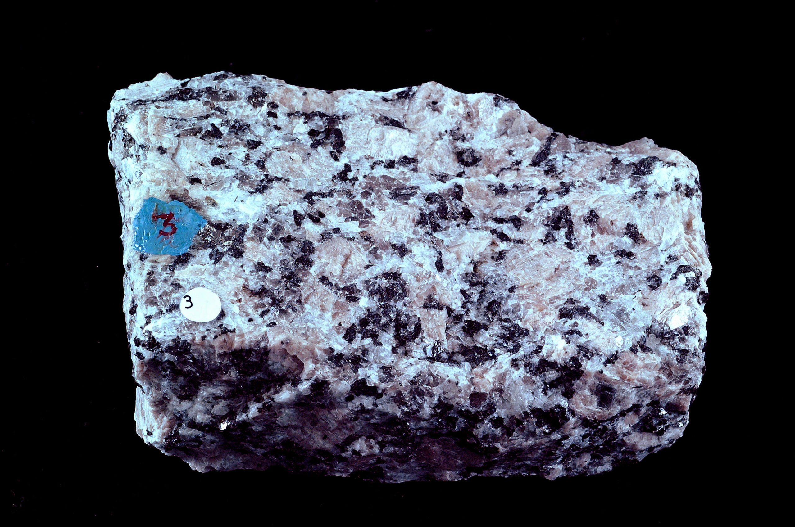
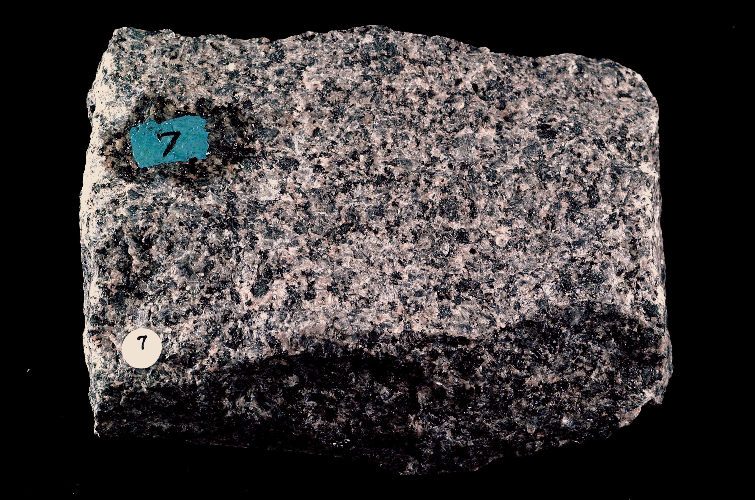
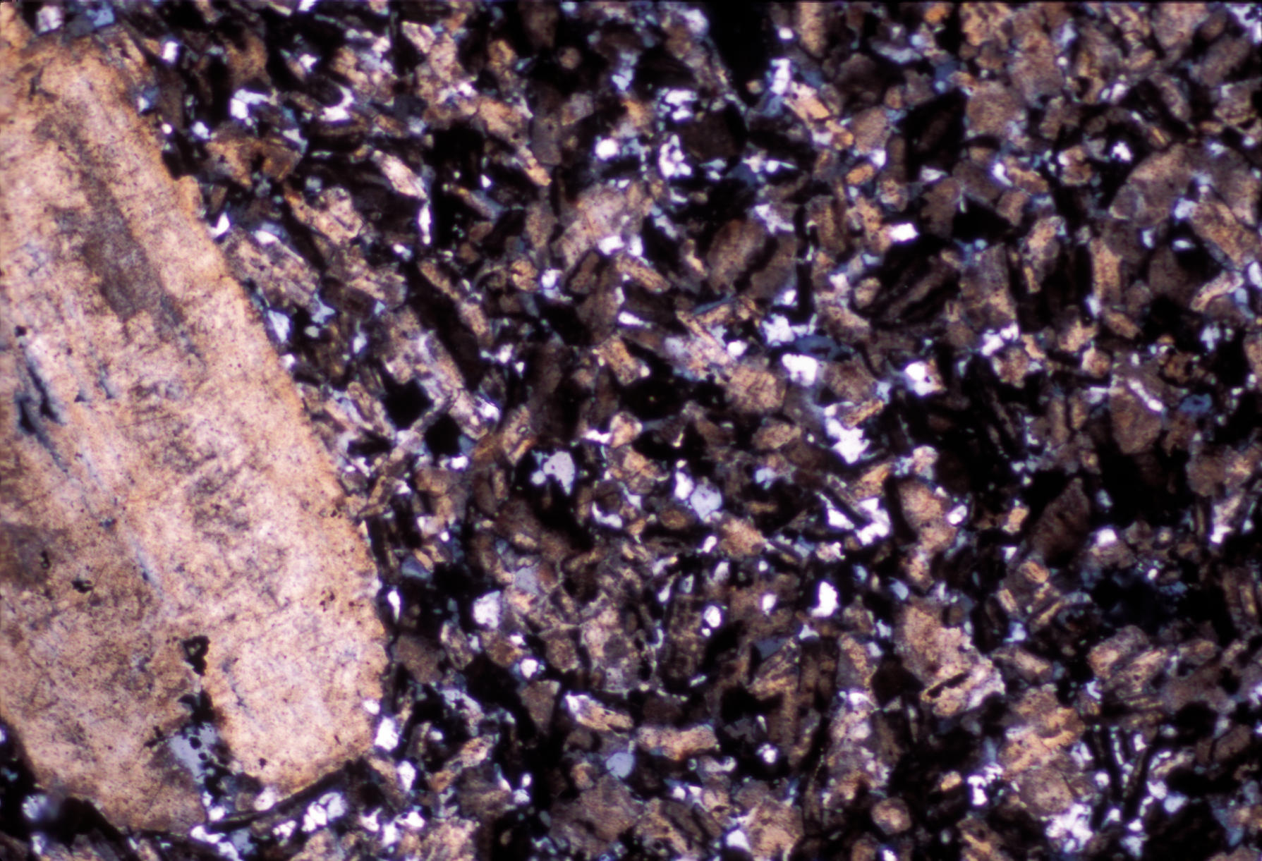
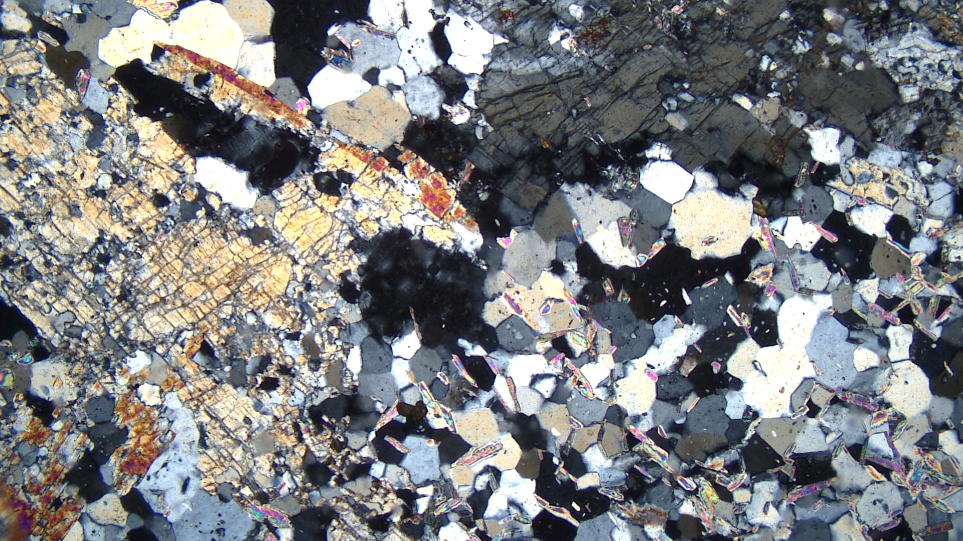
Granites are rocks generally dominated by quartz and K-feldspar. The coarse-grained granite seen above comes from St. Cloud, Minnesota. Quarries near St. Cloud are well known as sources of building stone, generically called the St. Cloud granite, whether the stone is truly granite or not. The St. Cloud granite is porphyritic, containing conspicuous large pinkish microcline crystals up to 1-2 cm across. Other light-colored mineral grains are gray plagioclase and glassy quartz. The darker colored minerals are biotite and hornblende but are hard to tell apart in the hand-sample view.
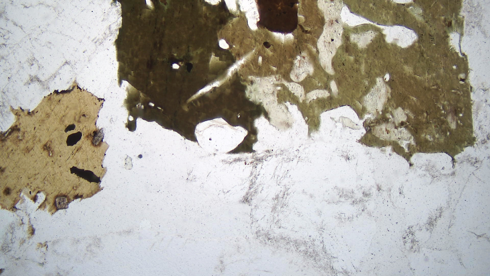
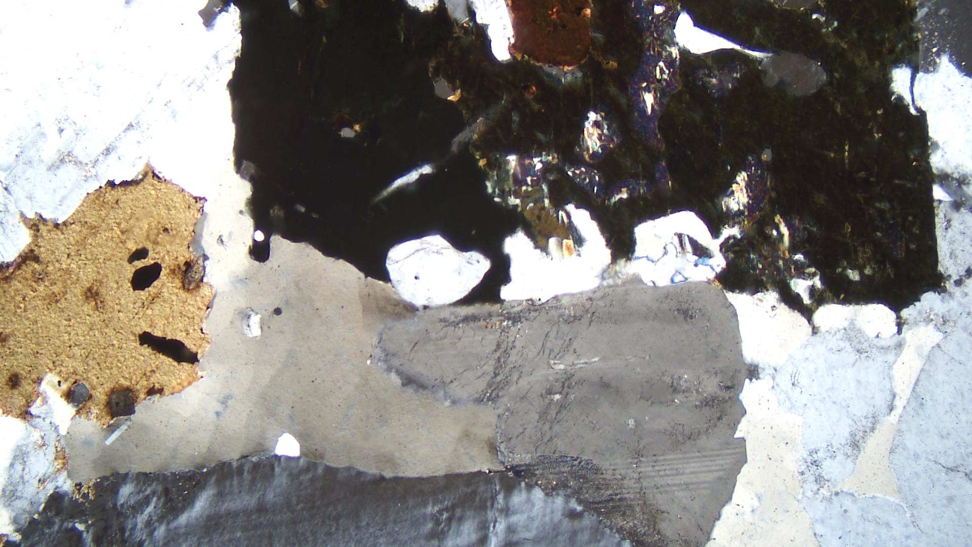
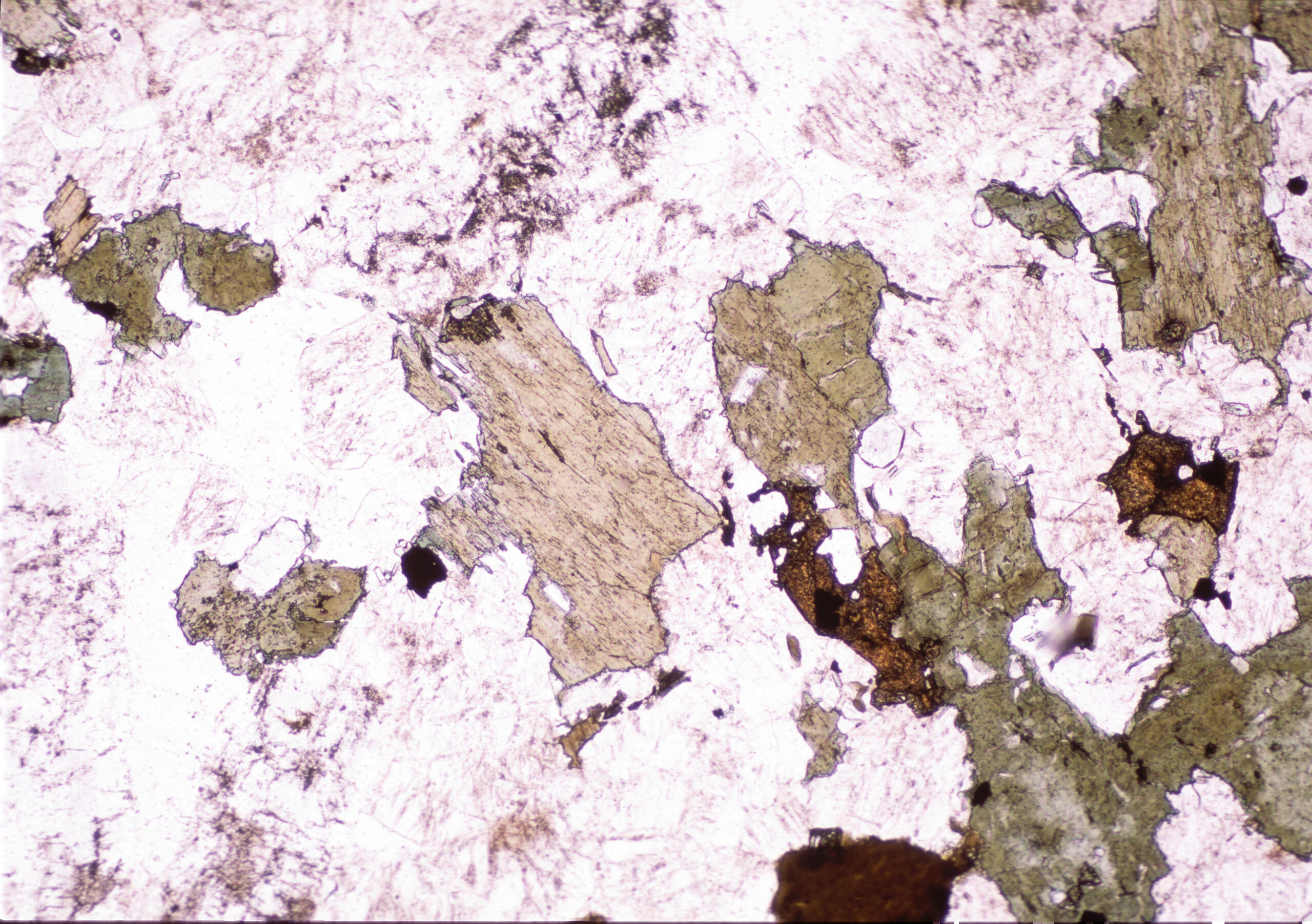
The PP thin-section view shows light brown flakes of biotite with green hornblende. Clear quartz and feldspar surround them. Both mafic minerals contain inclusions of ilmenite, and the larger biotite flake also has inclusions of high-relief titanite. In crossed polars, the interference colors of the mafic minerals do not show – being masked by the minerals’ strong colors. The large grain along the bottom edge of the view is K-feldspar, and just above it is a large quartz crystal displaying undulatory extinction. The other clear grains are all quartz or K-feldspar.
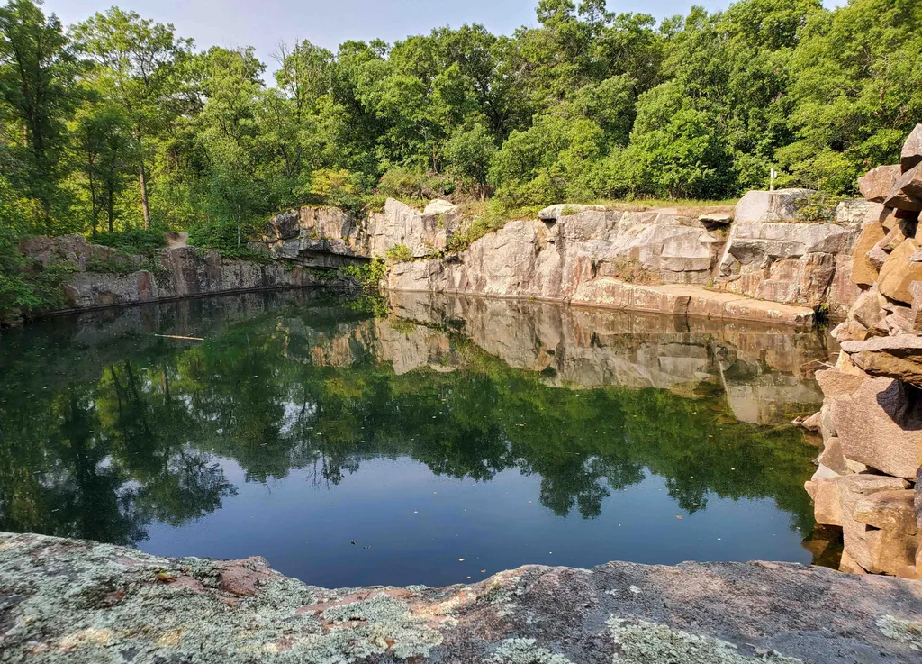
Figure 16.8 is a photo of one of the many abandoned quarry pits in the St. Cloud area.
▪Granodiorite
|
|
|
|

| ⇒Link to 3D rotatable image of the St. Cloud granodiorite |
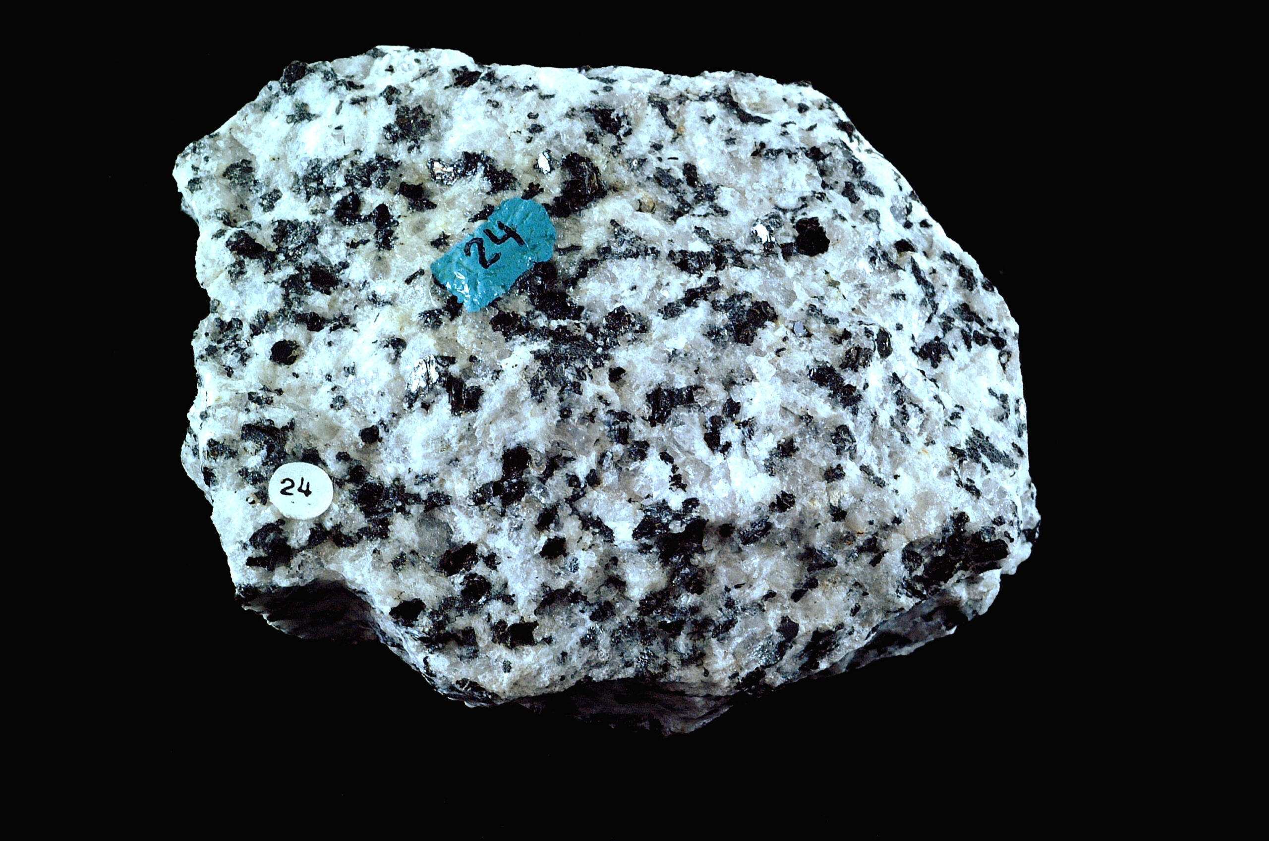
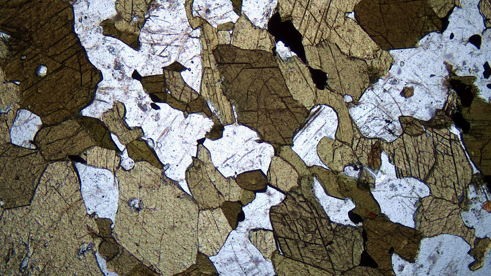
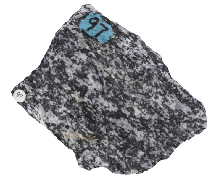
This granodiorite is another rock from near St. Cloud, Minnesota. It is mostly pink K-feldspar, gray euhedral to subhedral plagioclase, and black biotite. These minerals can be seen in the hand-sample photo. Lesser amounts of hornblende and quartz are also present but difficult to discern.
In plain-polarized light, the thin section shows brown biotite and pale-green hornblende. Somewhat altered feldspars and clear quartz are also seen. Two very high-relief dark-brown titanite crystals stand out. In some parts of this thin section, the hornblende contains remnants of augite, although it is not obvious in this view. In crossed polars, K-feldspar can be seen to be perthitic. Biotite displays 2nd-order colors. Plagioclase is zoned and a bit altered to sericite, and titanite displays its normal very high birefringence.
▪Tonalite
|
|
|
|


| ⇒Link to 3D rotatable image of this tonalite |
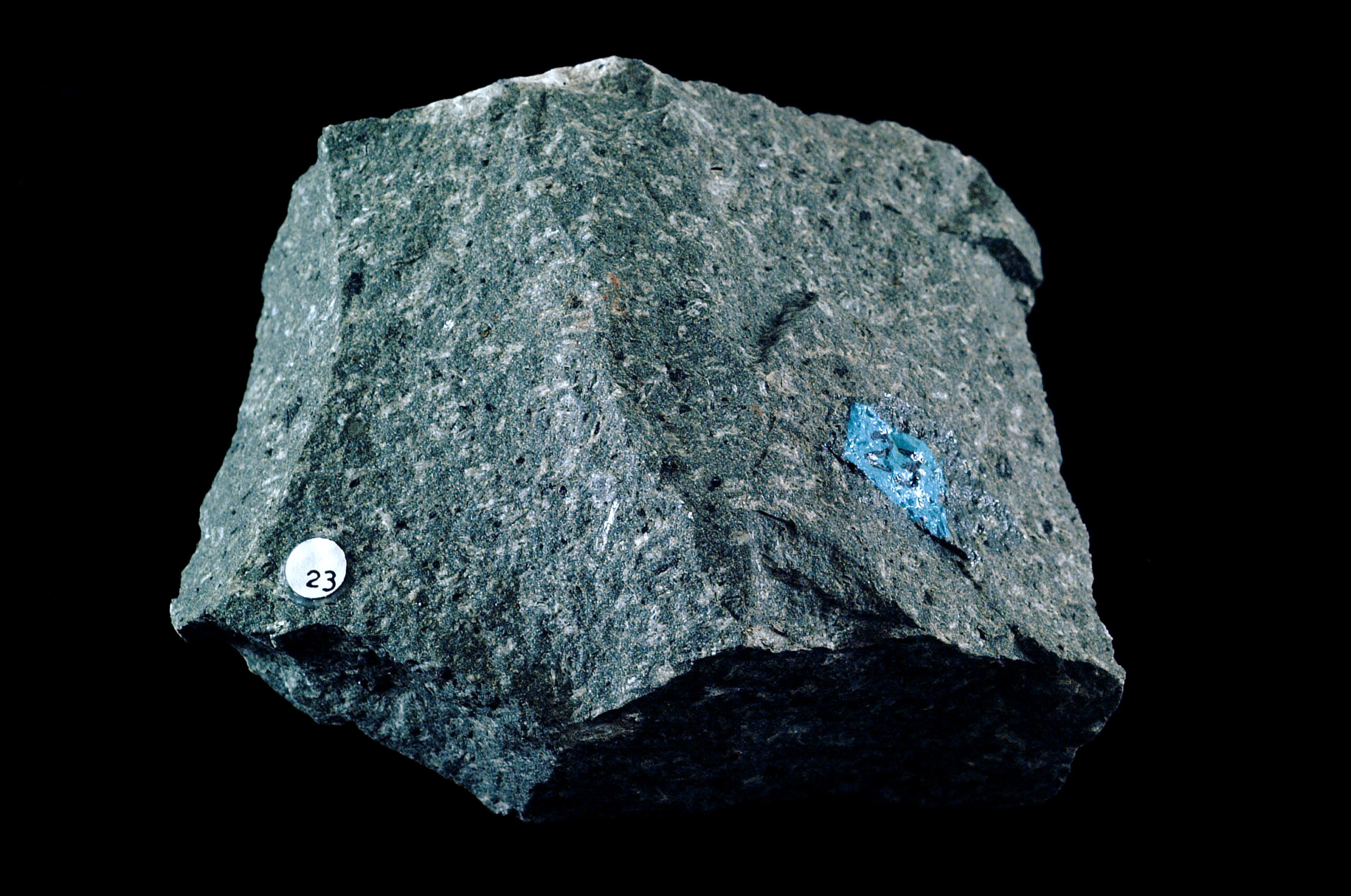
Tonalites are plutonic rocks characterized by essential quartz and plagioclase. K-feldspar may be present but only in small amounts. Mafic accessory minerals include biotite, hornblende, and clinopyroxene. The photos above are the Bonsall Tonalite from San Diego County, California. It contains primarily white plagioclase, gray quartz, and black biotite, all easily seen in the hand specimen.
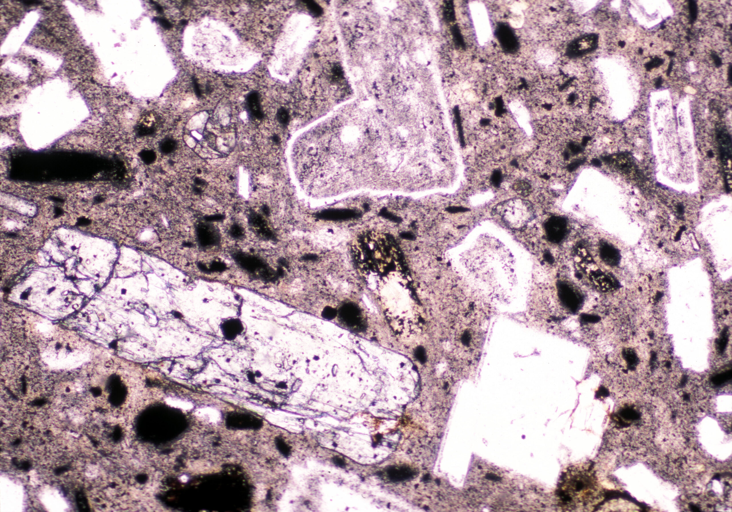
In the PP thin-section view, clear grains are quartz and plagioclase. Plagioclase has slightly higher relief than quartz and shows specks and lines due to alteration. Green hornblende and brown biotite are also present.
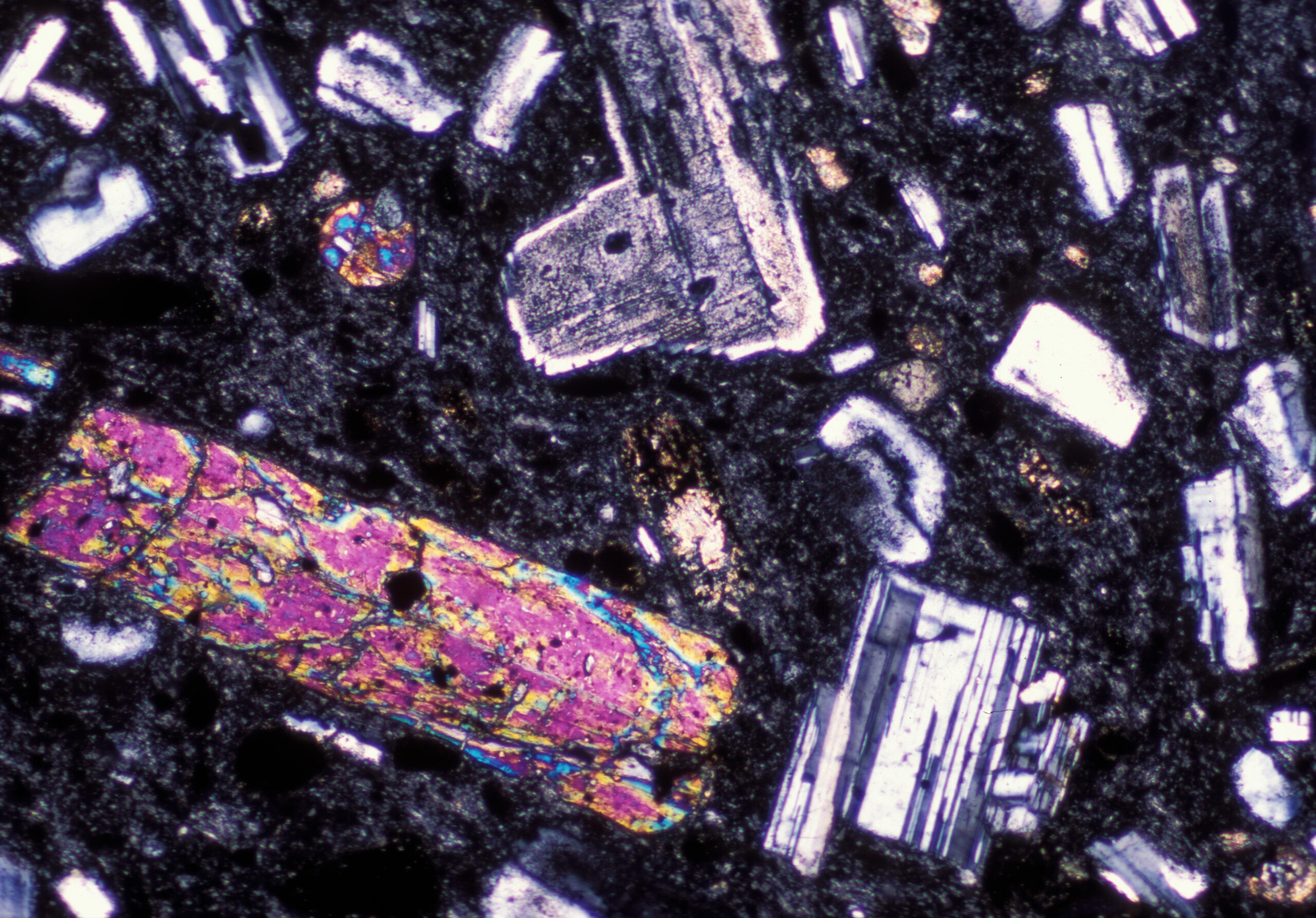
In the XP view, the diagonal lath of hornblende displays twinning and up to high 2nd-order interference colors. Biotite has its typical pebbly texture with up to 2nd-order colors. Plagioclase twins are evident but not as sharp as in many rocks.
16.1.2 Quartz-Poor Granitoids
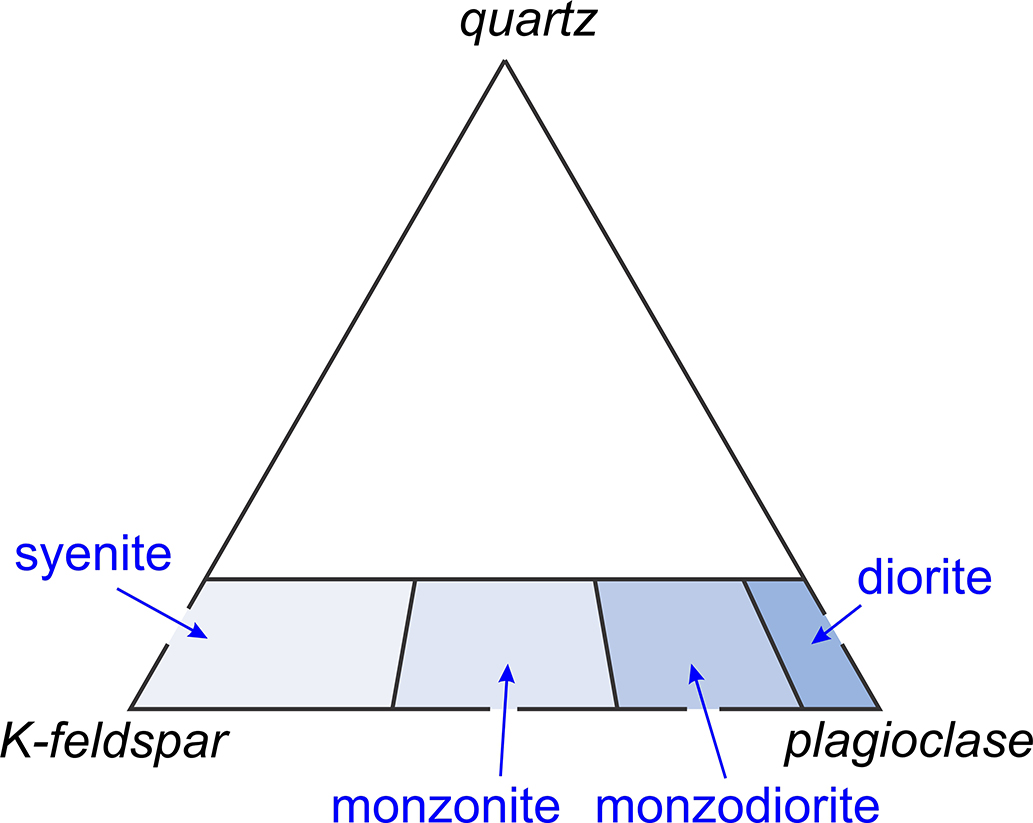
Quartz-poor granitoids are not as common as their quartz-rich cousins. These rocks contain 0-20% quartz with K-feldspar, plagioclase, or both. They range from syenites, which are rich in K-feldspar, to diorites, which are rich in plagioclase (Figure 16.15). As with other granitoids, there are many variations because of different minor and accessory minerals that may be present. Micas, amphiboles, and pyroxenes of various sorts, and other minerals, are common accessories.
▪Syenite
|
|
|
|

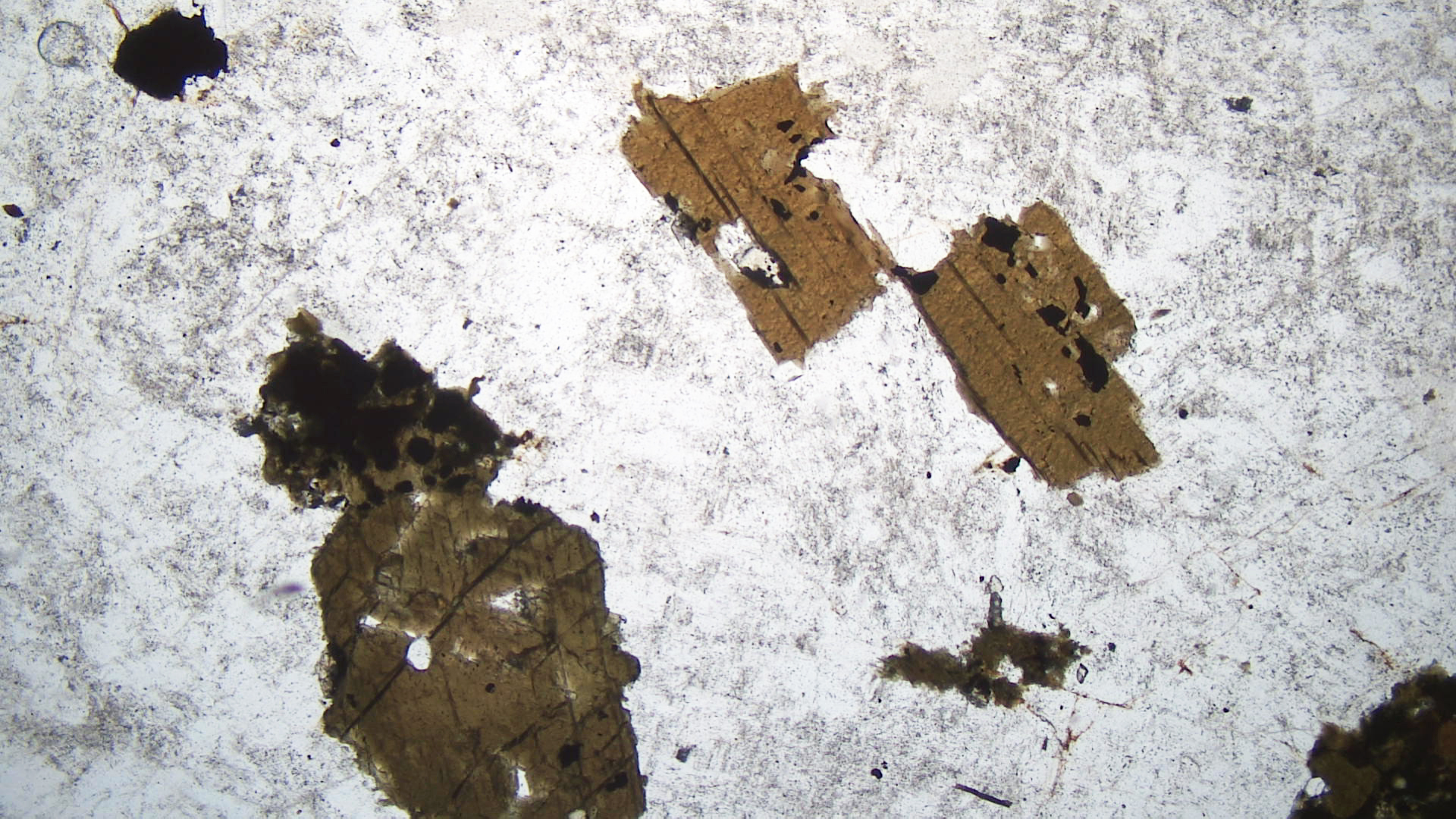
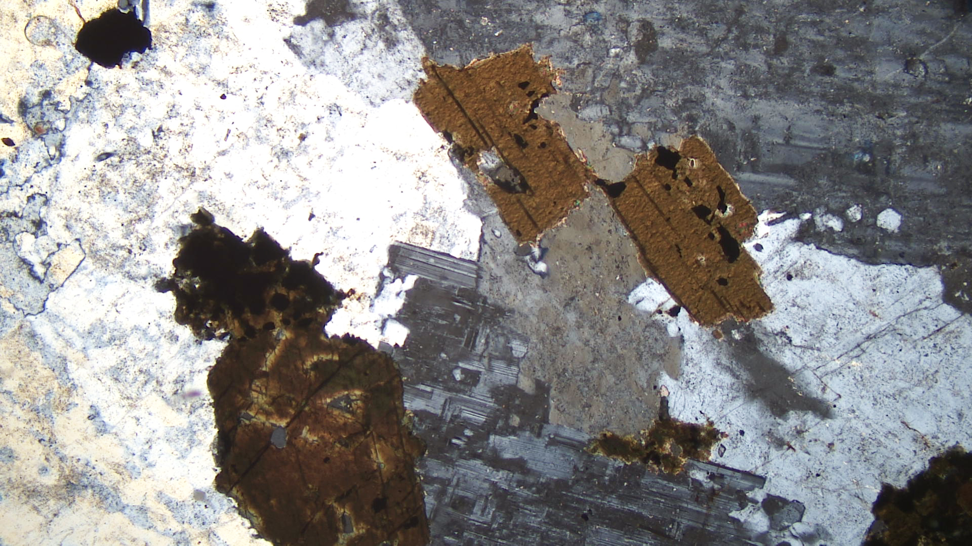
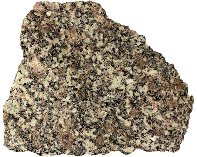
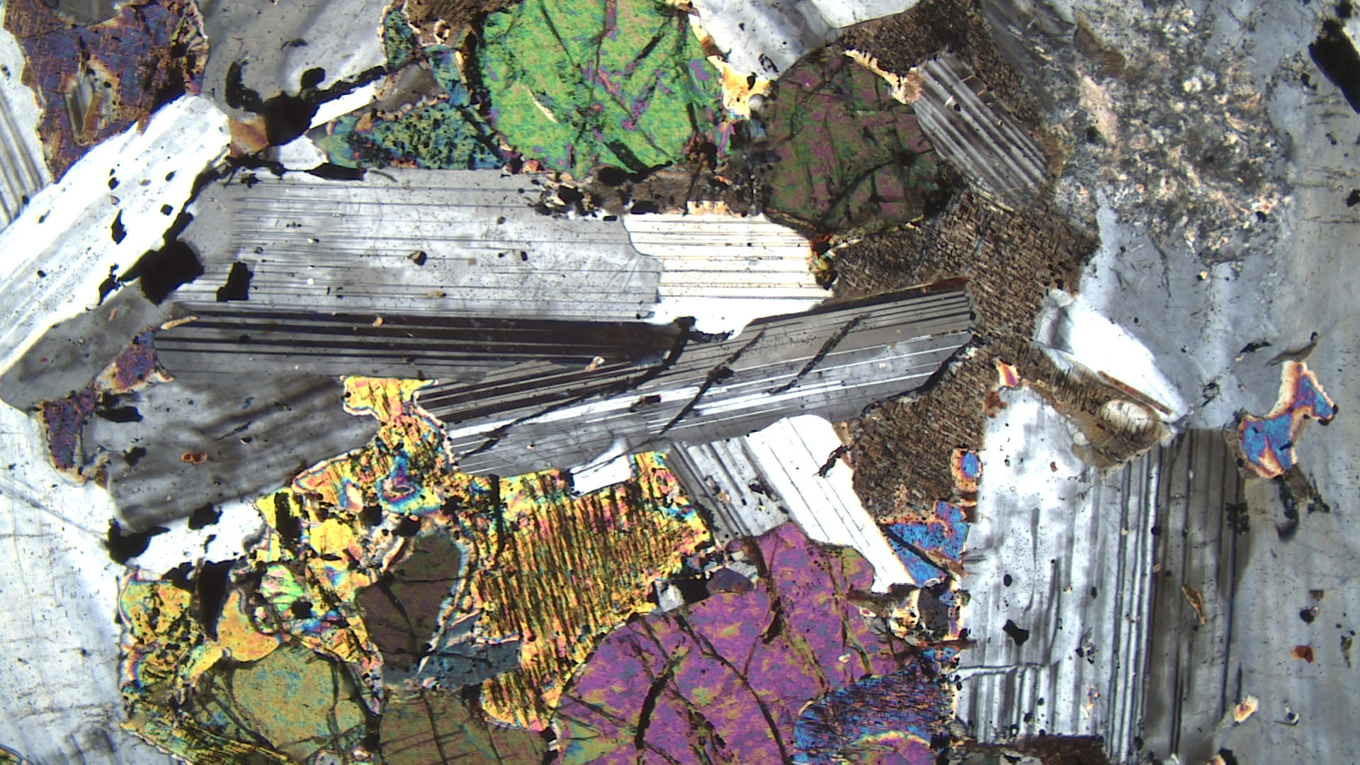
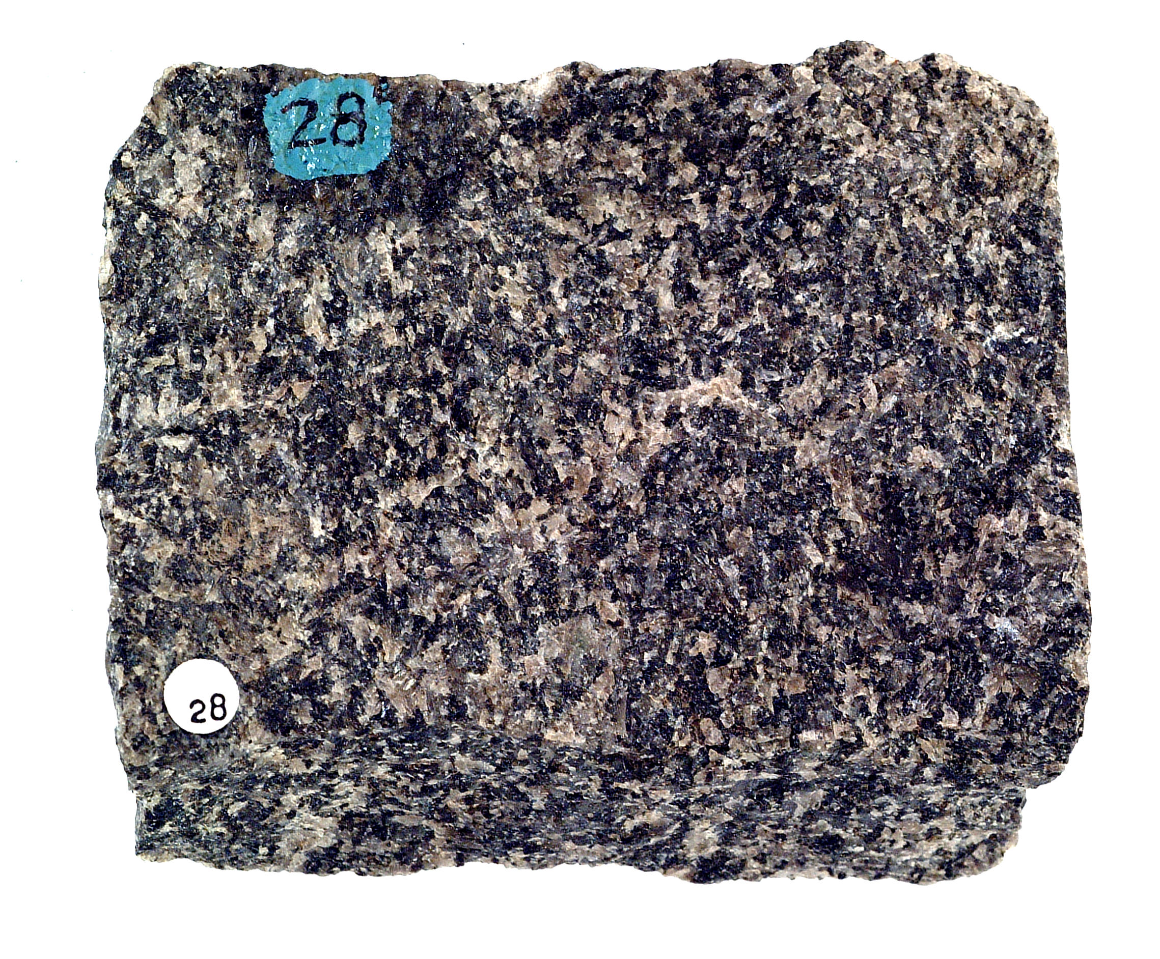
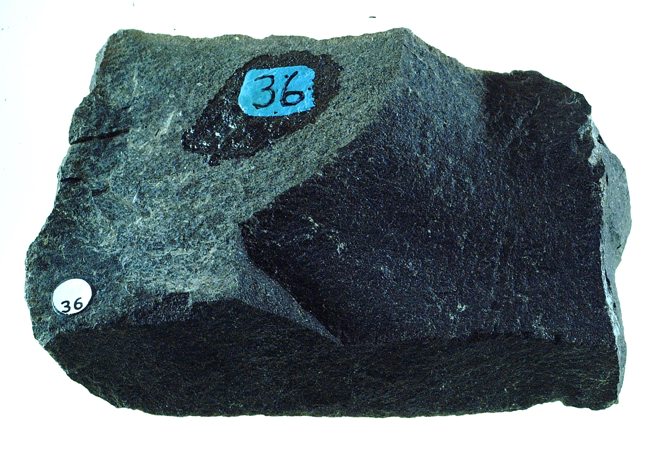
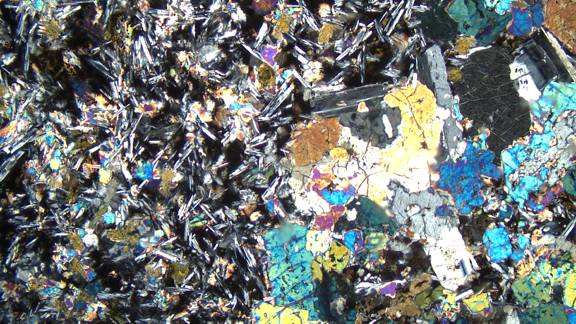
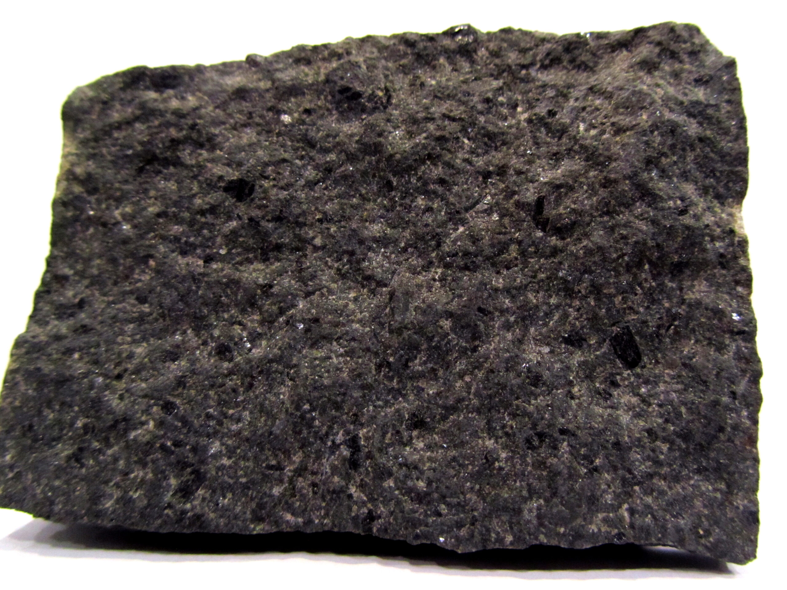
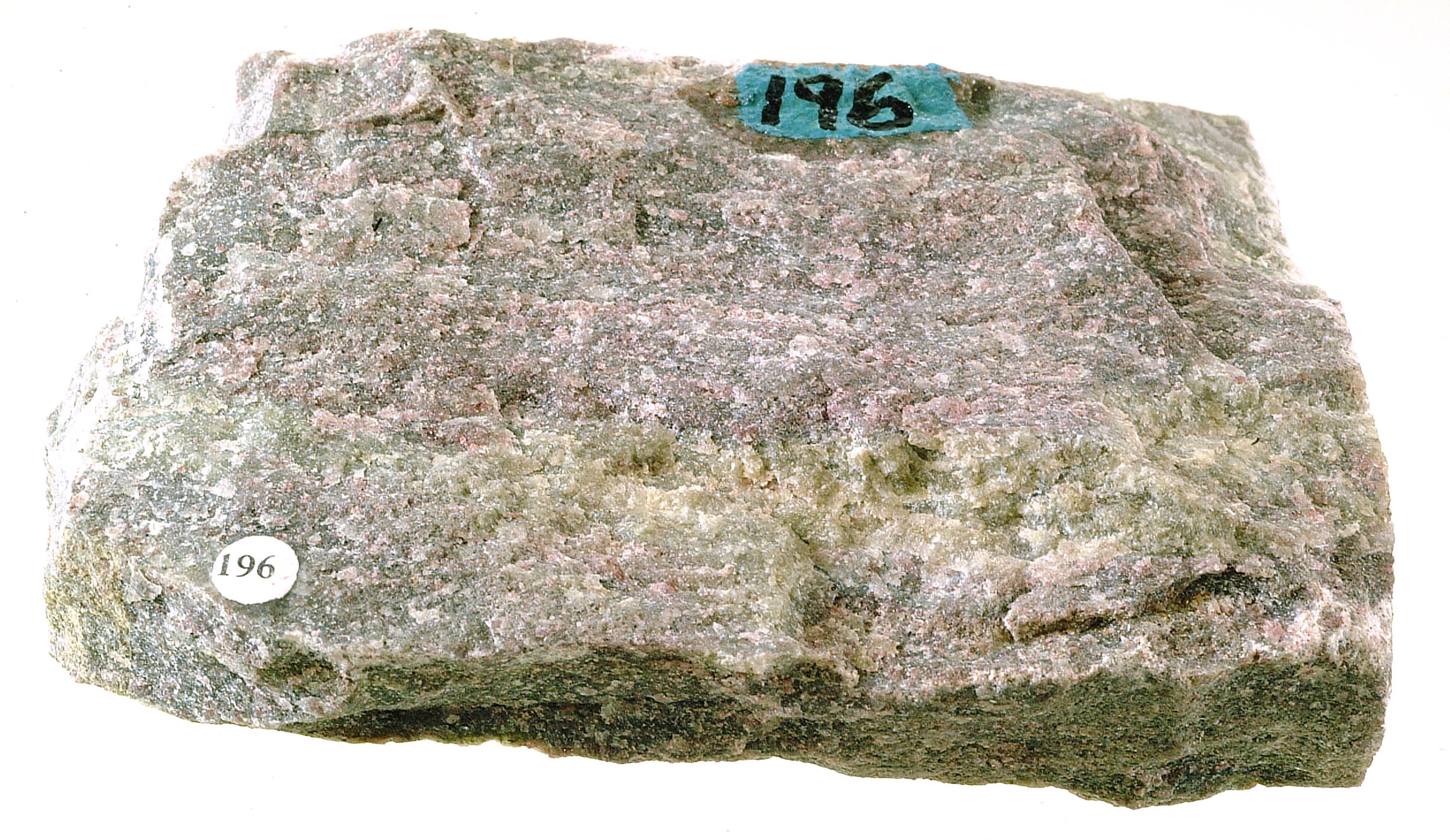
Syenites are rocks similar to granites but deficient in quartz. The coarse-grained syenite from Cuttingsville, Vermont (Figure 16.16) contains mostly light-colored perthitic orthoclase and Na-rich plagioclase. The mafic minerals are biotite with lesser amounts of hornblende. Minor quartz is present. Most of the dark minerals seen in the hand sample are biotite.
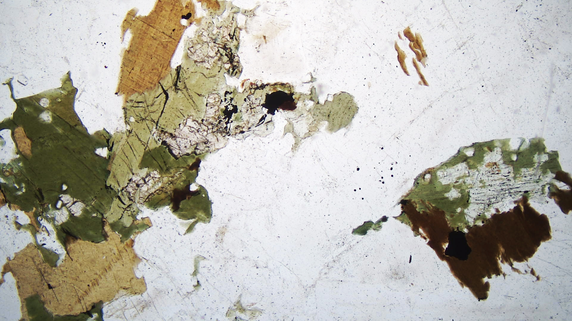
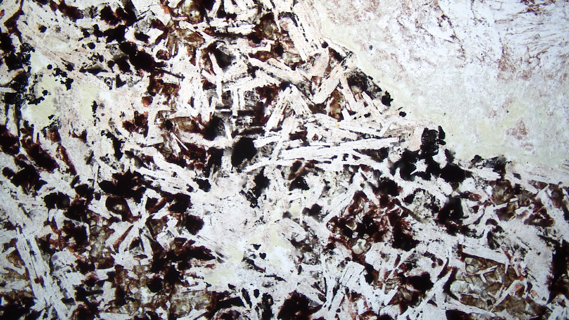
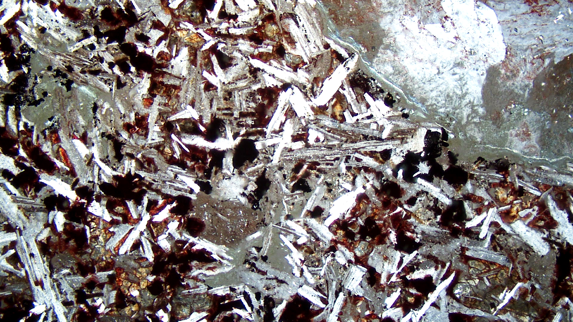
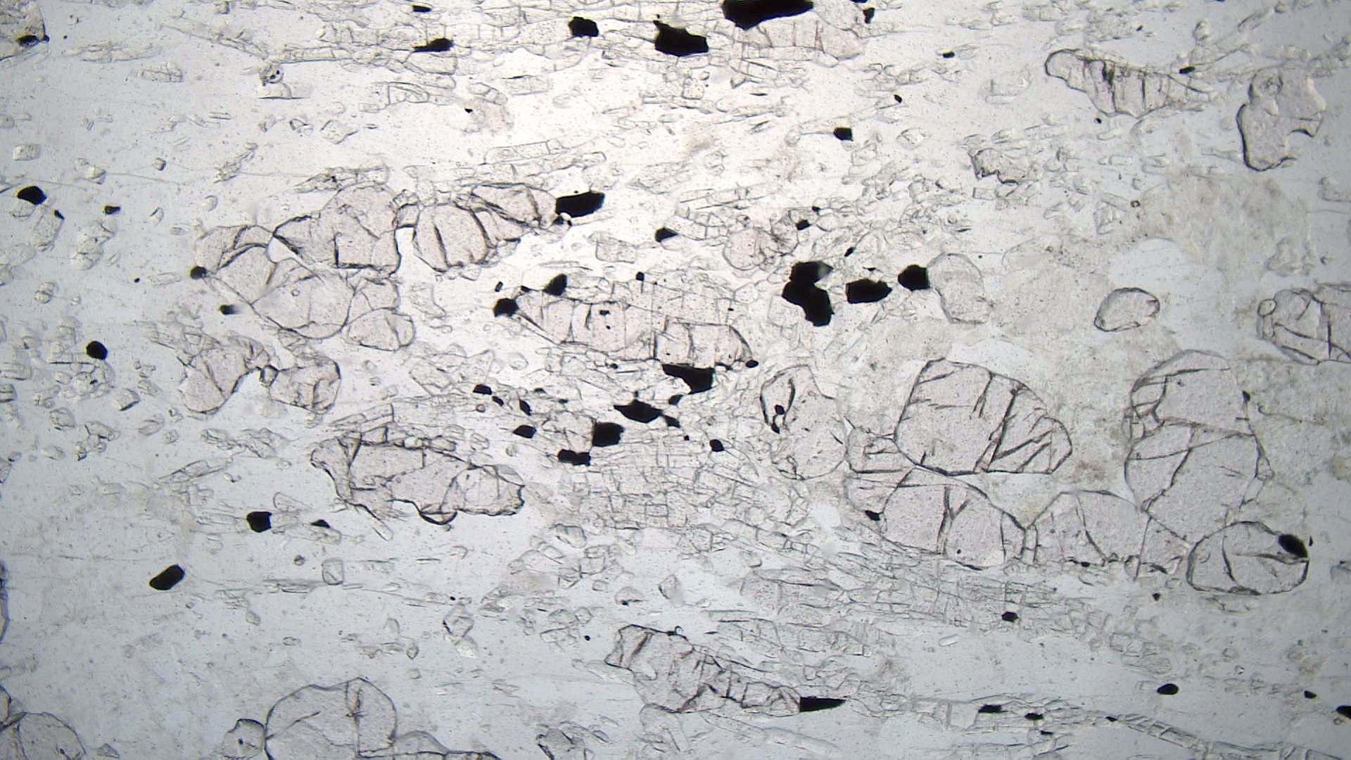
The thin-section views are of a different syenite. The PP view contains two flakes of brown biotite (upper part of view, right of center) with well-developed cleavage in one direction. The biotite contains inclusions of ilmenite. Below and to the left of the biotite is a large grain of greenish-brown hornblende that shows 60o-120o cleavage angles. The rough-textured clear grains are slightly altered feldspar.
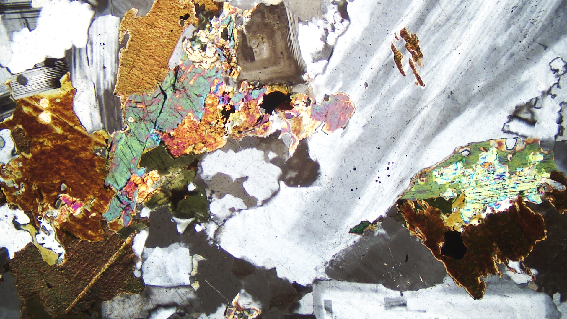
Under crossed polars, both mafic minerals appear brown due to the strong coloration of the mineral grains. Feldspars and quartz have their characteristic black-gray-white interference colors. A large grain of plagioclase displays twinning, but most of the feldspar grains are K-feldspar.
▪Monzonite
|
|
|
|
| ⇒Link to 3D rotatable image of a different monzonite |
Monzonites are plutonic rocks that contain subequal amounts of plagioclase and K-feldspar, and minor amounts of quartz. The hand sample of monzonite above is from an unknown location in France. Besides the two feldspars, it contains a small amount of mafic minerals. In the hand-sample view, the K-feldspar is pink, the plagioclase is whitish, and the mafics appear black.
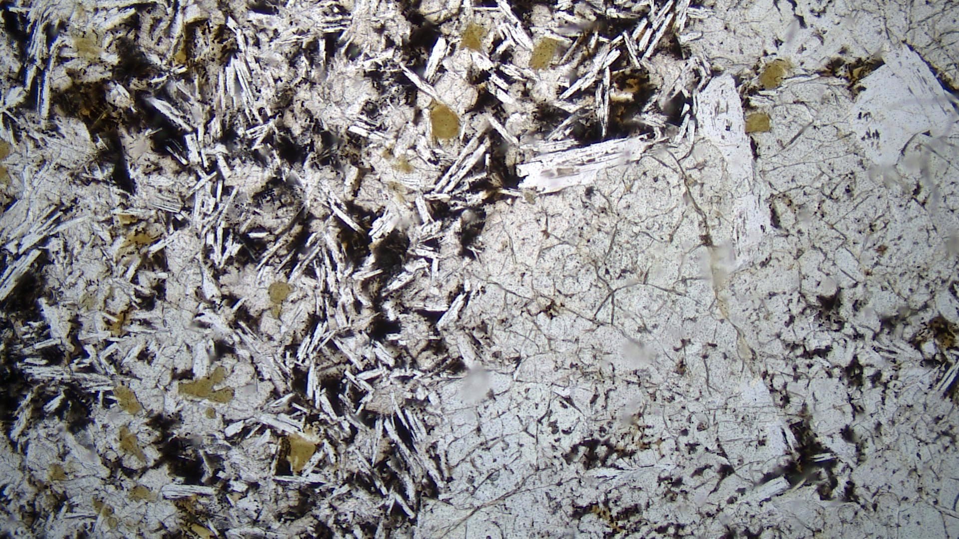
The thin sections views above are of a different monzonite, one from the Monzoni Massif in Italy, the type locality for this kind of rock. The colorless grains in the PP view are orthoclase and plagioclase. Green hornblende and relatively high-relief pale gray-green to pink clinopyroxene are also present.
In crossed polars, the pyroxene has strong 2nd-order or 3rd-order interference colors. It is hard to see hornblende’s interference colors because the strong color of the mineral masks them. Plagioclase grains are smaller than the K-feldspars. Plagioclase shows polysynthetic twinning. Some K-feldspar grains show simple Carlsbad twinning.
▪Quartz Monzonite
|
|
|
|


| ⇒Link to 3D rotatable image of this quartz monzonite |
Quartz monzonites contain quartz and subequal amounts of plagioclase and K-feldspar. These rocks may be porphyritic, and biotite or hornblende are typical accessory minerals. This quartz monzonite from Garfield, Colorado, contains phenocrysts of very light-pinkish K-feldspar and white plagioclase in about equal proportions. The two feldspars have about the same color in the hand sample. Quartz crystals are gray. The mafic minerals are biotite and hornblende. The rock contains 10-25% quartz.
In the plain-polarized view, the clear grains are plagioclase, orthoclase, and minor quartz. The quartz grains are clearer than the feldspar grains because they have not altered. A pale green hornblende lath runs diagonally across the view. The lath contains some small included grains of high-relief epidote. A large grain of biotite near the left side of the photo has altered to hornblende along its edges. Minor magnetite (opaque) is also present.
Twinned plagioclase dominates the upper-right part of the XP view. An altered grain of orthoclase is seen near the top just left of center. Some smaller quartz grains are along the bottom edge; they have no twinning and no alteration. Hornblende interference colors are up to middle 2nd order. Biotite’s interference colors are hard to make out but also reach 2nd order.
▪Diorite
The figures below are photos of a diorite from near Azusa, California.
|
|
|
|
| ⇒Link to 3D rotatable image of this diorite |
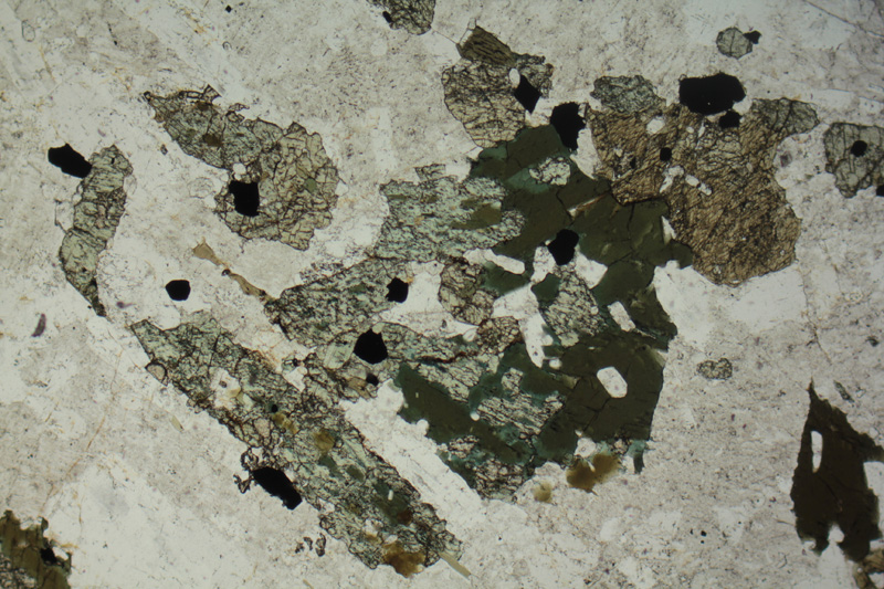
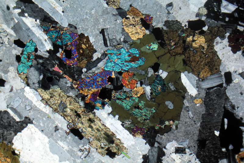
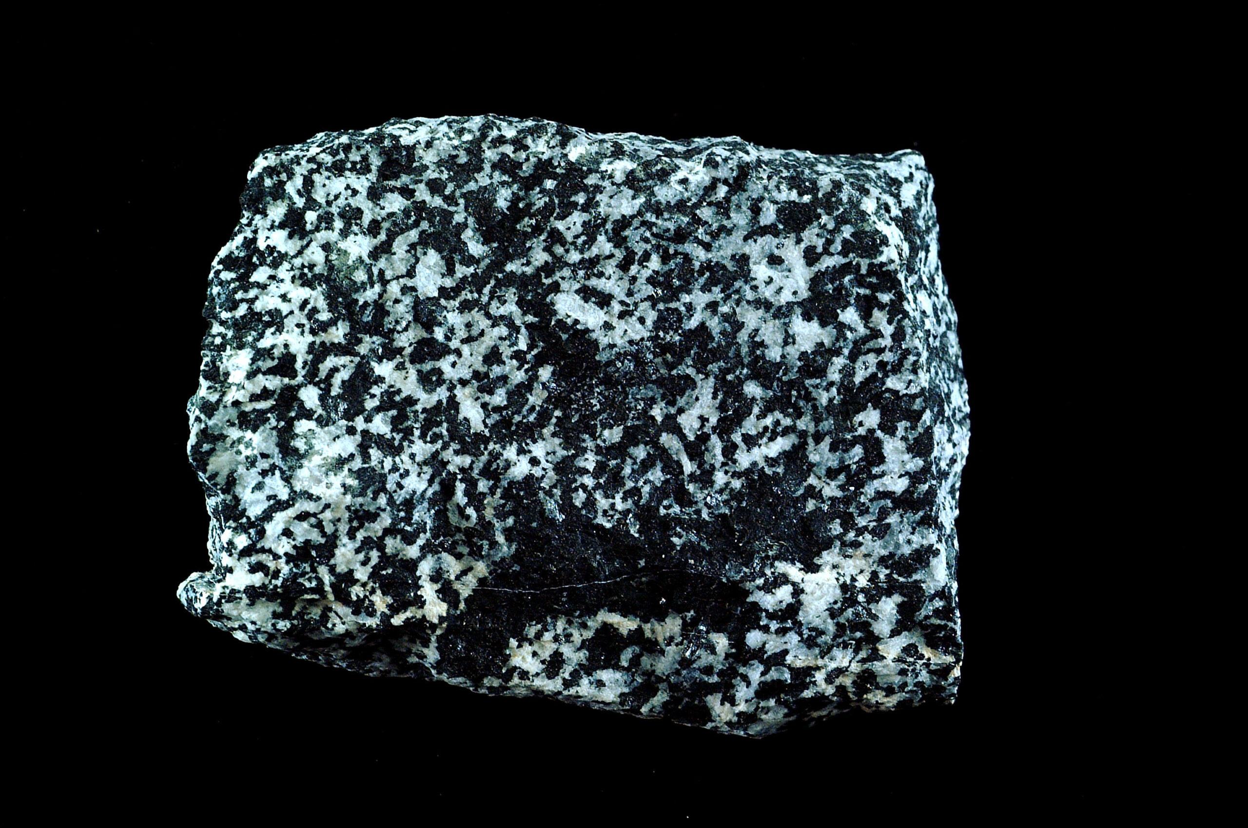
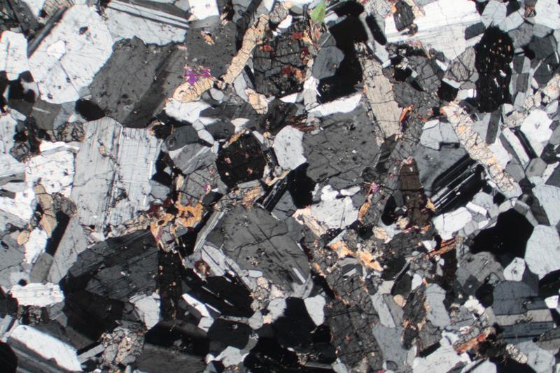
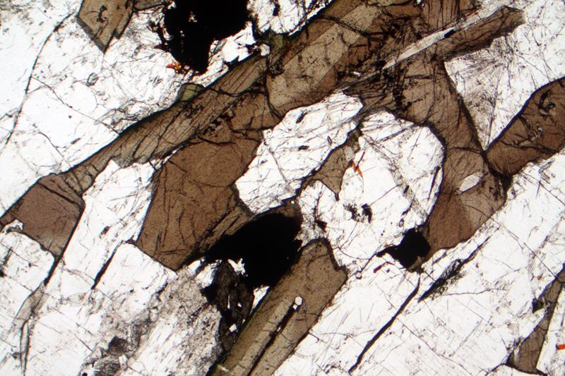
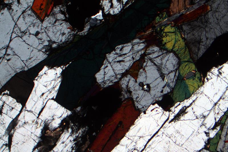
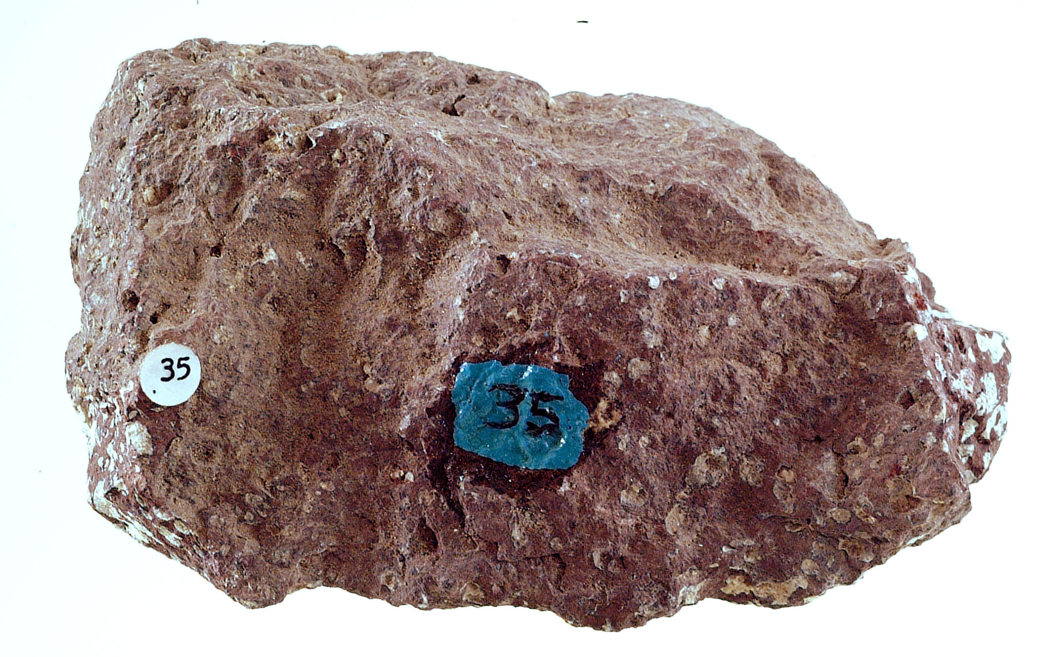
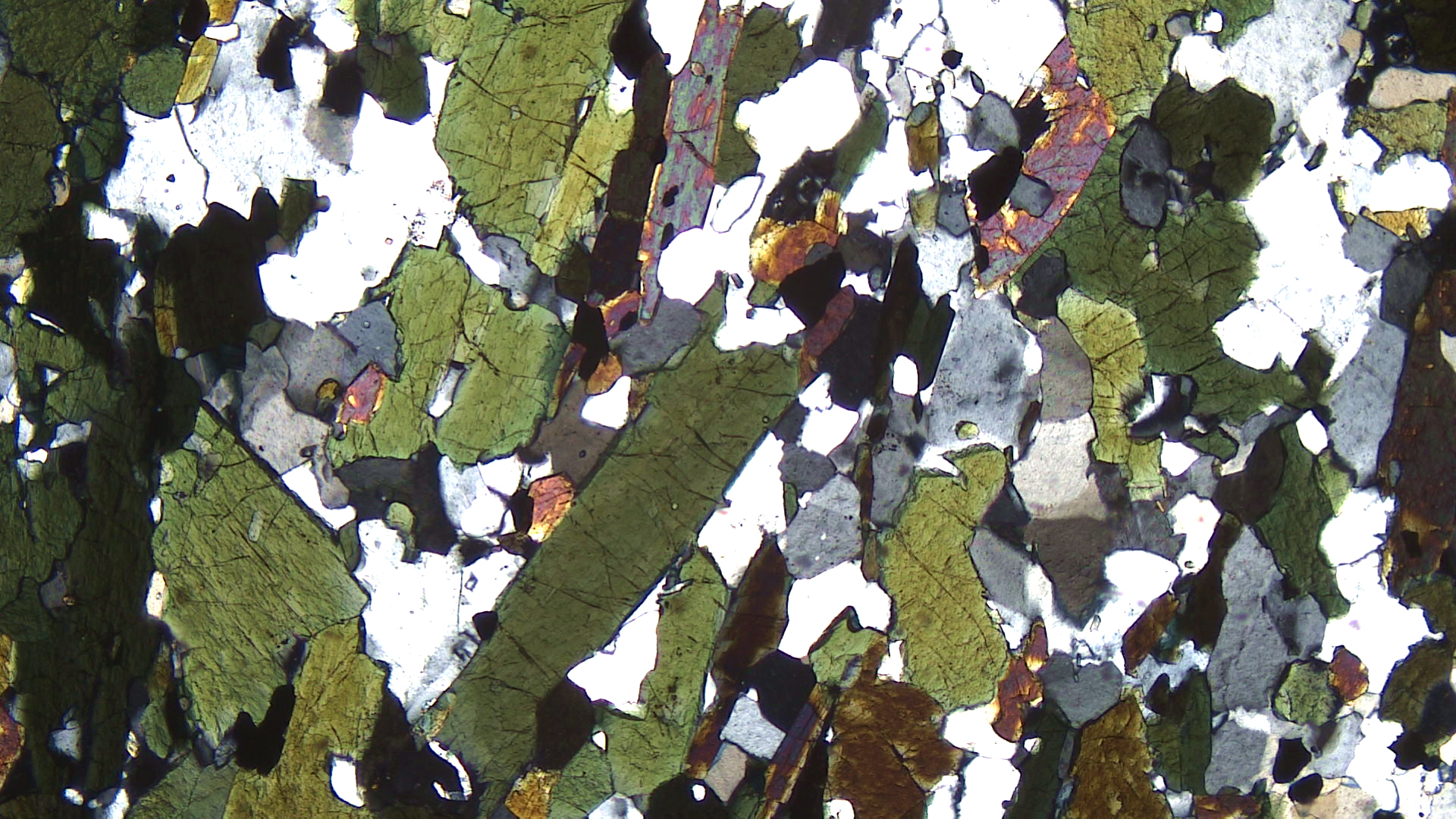
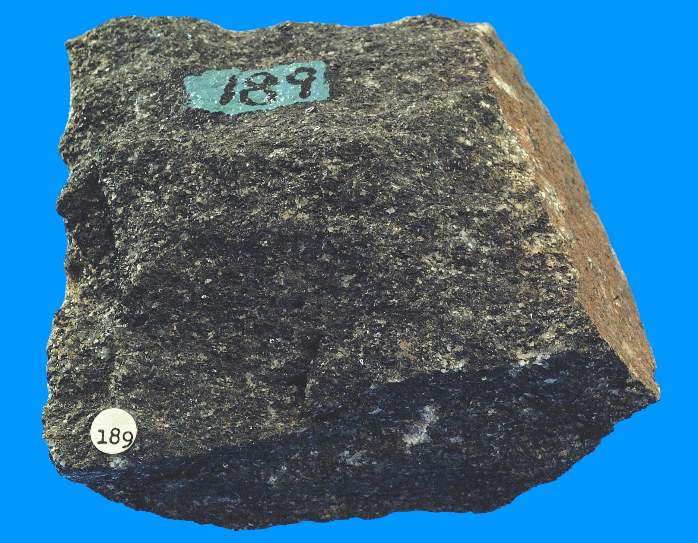
Diorites are mafic rocks dominated by plagioclase, amphiboles, and pyroxenes, sometimes with small amounts of biotite. The hand specimen above has the typical look of a diorite: a mix of black and white equal-sized grains. Sometimes we describe this as a “salt and pepper” appearance. The dark mineral is hornblende in this sample, and the white mineral is plagioclase. Most mineral grains are 1-4 mm in longest dimension, but a few hornblende crystals are significantly larger.
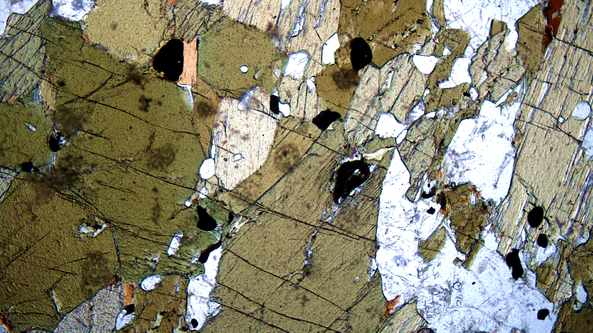
In thin section, the PP view shows mostly hornblende (in various shades of green) and plagioclase (clear). Minor biotite is mixed with the hornblende in a couple of places but is hard to pick out. Some opaque mineral, probably magnetite, is also present. In crossed polars, plagioclase has typical lamellar twinning. Hornblende has up to middle 2nd-order interference colors. The biotite crystals are too small to see interference colors.
16.1.3 Gabbroic Plutonic Rocks
The IUGS classification scheme seen in Figures 16.1 and 16.15 does not work well for rocks that contain only plagioclase and no K-feldspar or quartz. Such rocks, gabbroic rocks, may be of several different kinds. They are distinguished based on other minerals besides feldspars and quartz and so require a different scheme for naming them.
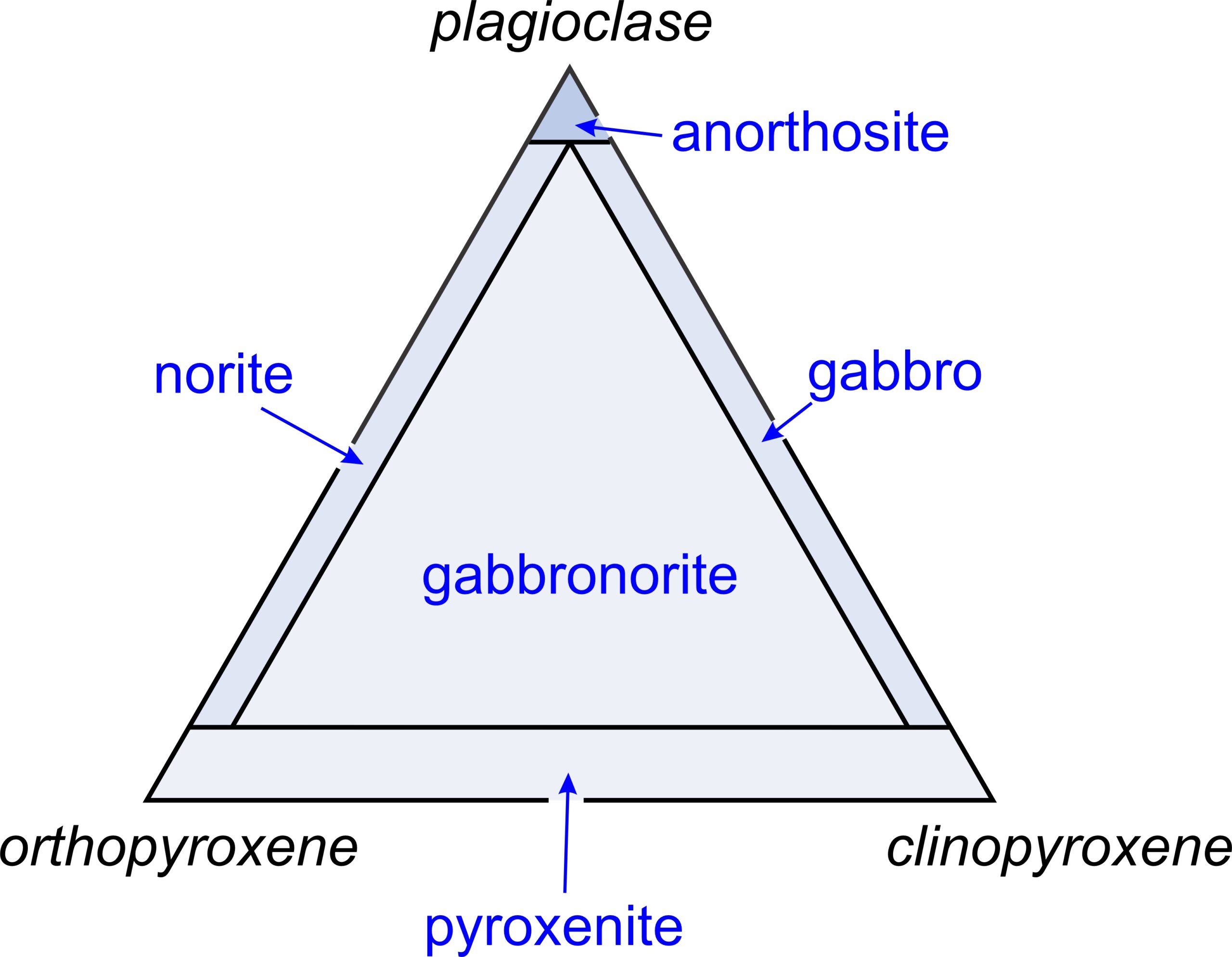
Figure 16.28 is the IUGS classification system for different kinds of gabbroic rocks. All rocks in this group contain some combination of plagioclase, orthopyroxene, and clinopyroxene. Common gabbros are intrusive equivalents of basalts and contain mostly plagioclase and clinopyroxene (augite). The gabbro group, however, covers a wider range of compositions. Gabbronorite is the generic name for rocks that contain significant amounts of all three minerals. As seen in the diagram, rocks primarily made of two of these minerals are given more specific names: gabbro, pyroxenite, or norite. Rocks composed nearly entirely of plagioclase are anorthosites. Olivine is a common accessory mineral in all rocks of the gabbro group. Troctolites, rocks composed primarily of olivine and plagioclase, are also considered members of the Gabbro group.
▪Gabbro

Gabbros in the Duluth, Minnesota, region are somewhat variable in mineralogy and texture. They are part of the 5,000 km2 Duluth Complex that contains many related rocks including gabbro, anorthosite, norite, and troctolite. Figure 16.29 is a photo of an outcrop at the Spirit Mountain ski area on Skyline Drive in Duluth. Pyroxene- and plagioclase-rich layers alternate. The layering developed because of crystal settling during crystallization.
blank
|
|
|
|

The hand specimen above is a sample of medium-grained gabbro from near Duluth. It contains, like all gabbros, blocky pyroxene (augite) crystal and laths of plagioclase that are a bit larger. Olivine, although present, is not easily seen.
The PP thin-section view above shows clear tabular plagioclase crystals and slightly darker colored, higher-relief, augite and olivine crystals. Some of the augite crystals contain dark twin lamellae that appear as fine parallel lines. Olivine crystals are blocky and fractured, and have slightly higher relief than the augite. Minor brown biotite and an opaque mineral are also seen.
In crossed polars, olivine displays variable and patchy 3rd-order interference colors. Augite’s interference colors are a bit lower. Plagioclase has typical zebra-stripe twins and, in places appears to be altering to a fine-grained clay or mica.
▪Hornblende Gabbro
|
|
|
|
| ⇒Link to 3D rotatable sample of this gabbro |
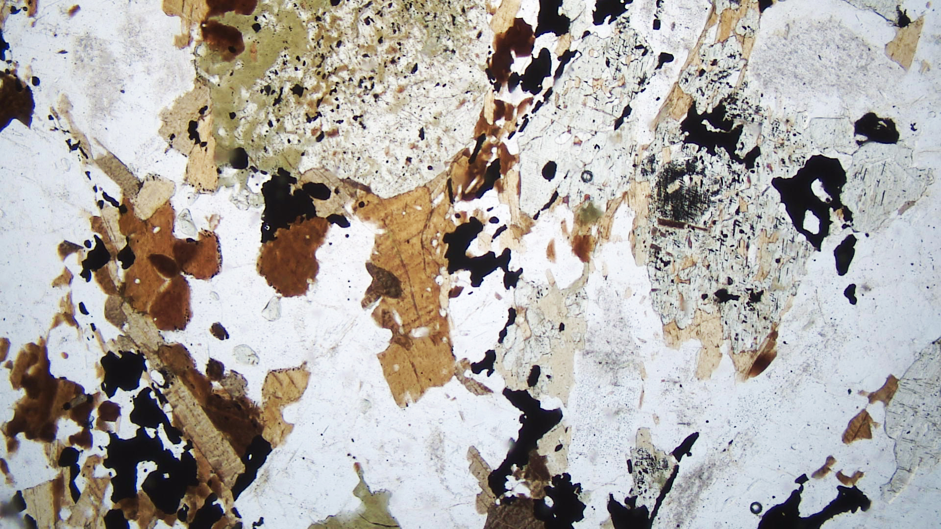
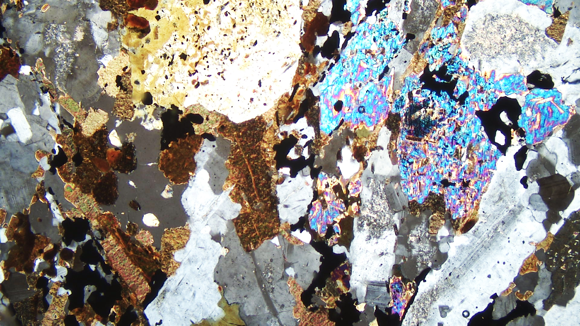
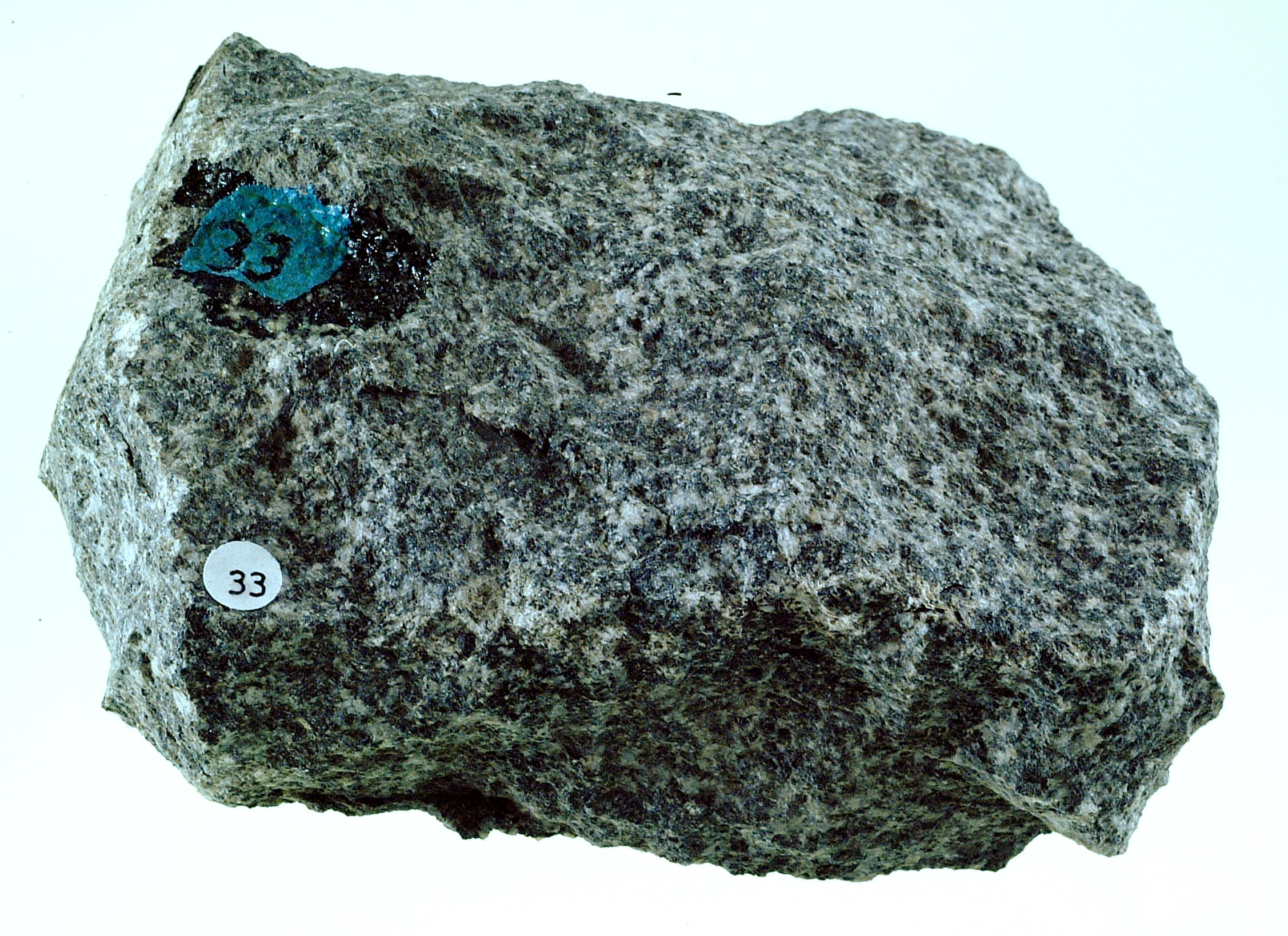
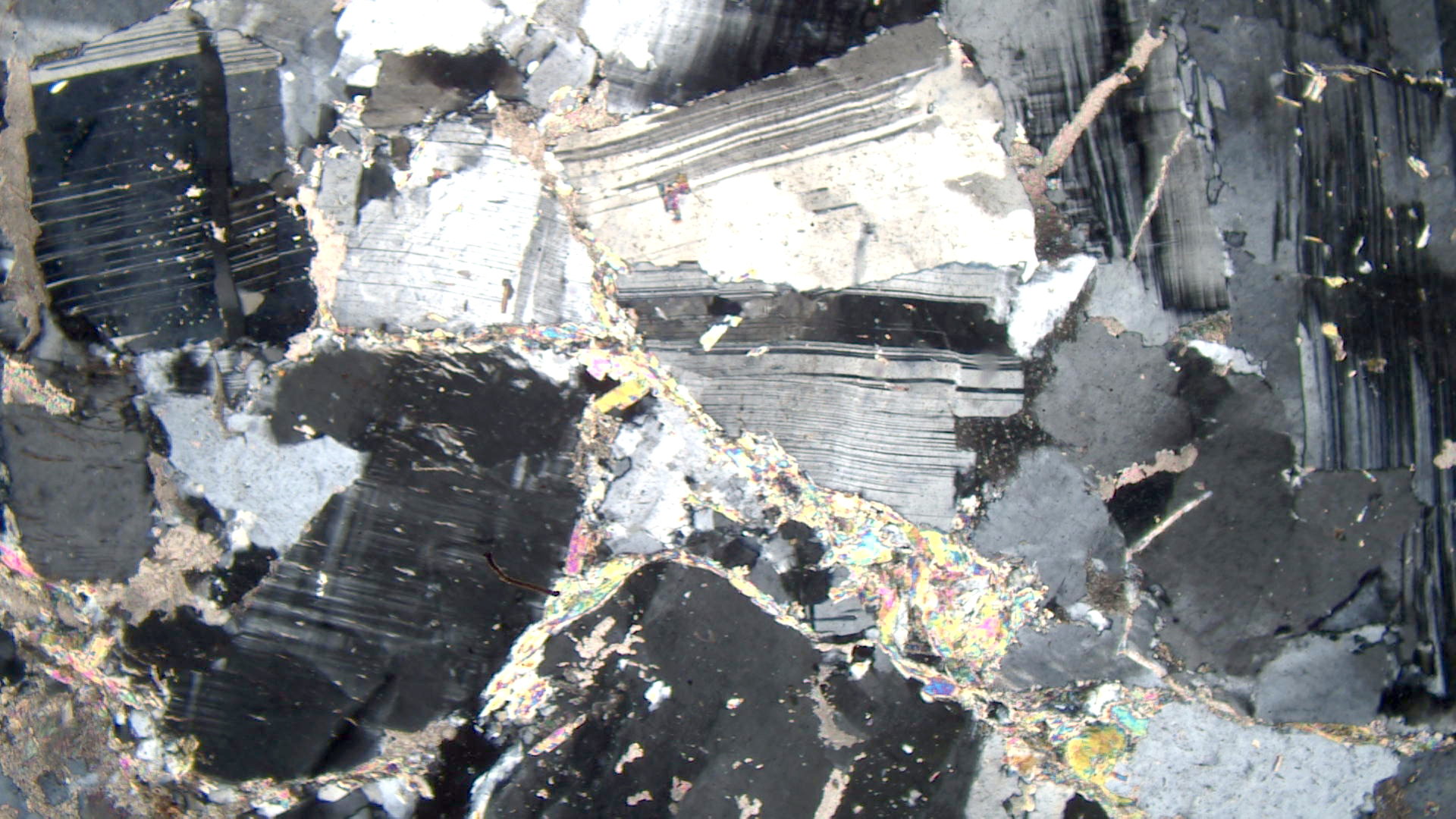
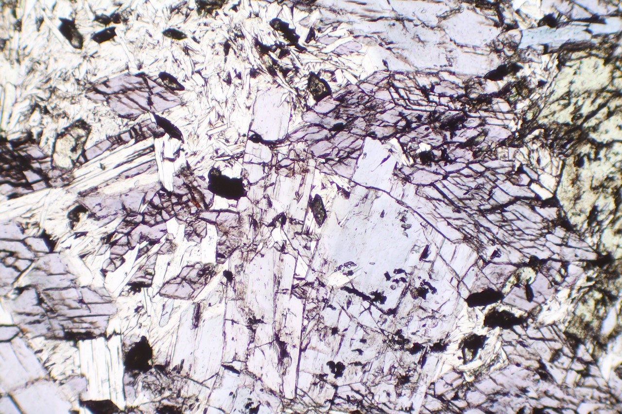
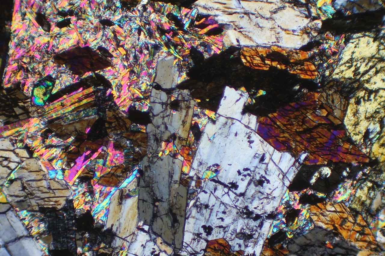
All gabbros contain clinopyroxene and plagioclase as primary minerals. However, this hornblende gabbro, from Escondido, California, contains mostly plagioclase and hornblende. The hornblende formed by alteration of original augite; some remnant clinopyroxene is still present. Plagioclase and hornblende crystals up to a centimeter long are somewhat aligned, giving the rock a slight foliation.
In the PP view, mafic minerals are seen to cluster. Brown biotite flakes are adjacent to very light-green augite that is altering to darker-green hornblende. Minor magnetite is present as well.
In crossed polars, augite displays up to 3rd-order interference colors, and biotite’s strong brown color masks its interference colors. Hornblende shows mostly 2nd-order, but variable, interference colors. Plagioclase is zoned and displays twinning.
▪Anorthosite
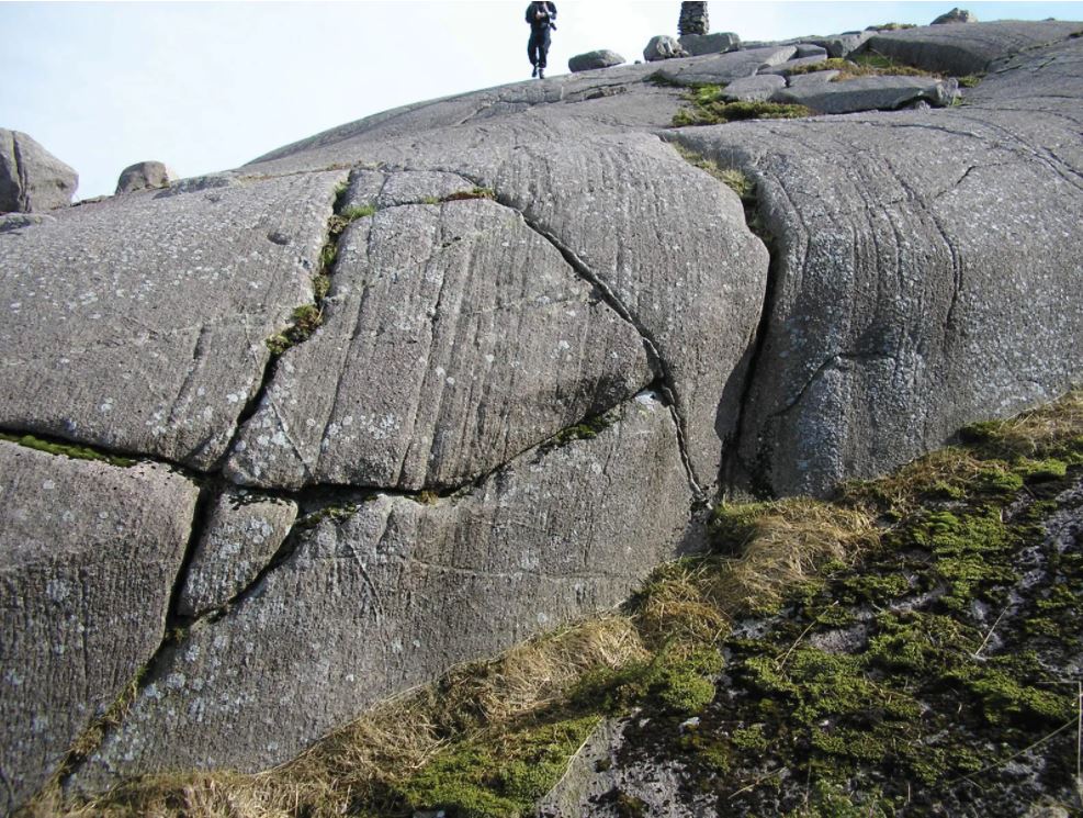
Anorthosites are relatively rare plutonic rocks consisting mostly of plagioclase (usually labradorite or bytownite). Small amounts of mafic and oxide minerals may be present as accessories.
Many anorthosite bodies are cumulates associated with other mafic rocks in layered complexes. The most common anorthosites, however, are in Proterozoic massifs. Figure 16.36 is a photo of an outcrop of anorthosite in the Helleren Massif of southwestern Norway.
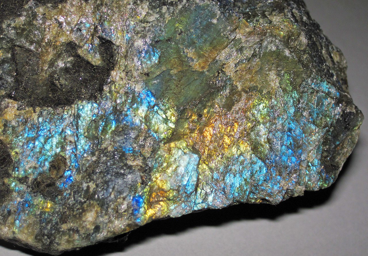
Some anorthosite contains labradorite which shows a remarkable blue schiller when polished. Figure 16.38 below shows an example. This rock is prized as a building stone.
|
|
|
|

| ⇒Link to 3D rotatable image of this anorthosite specimen |
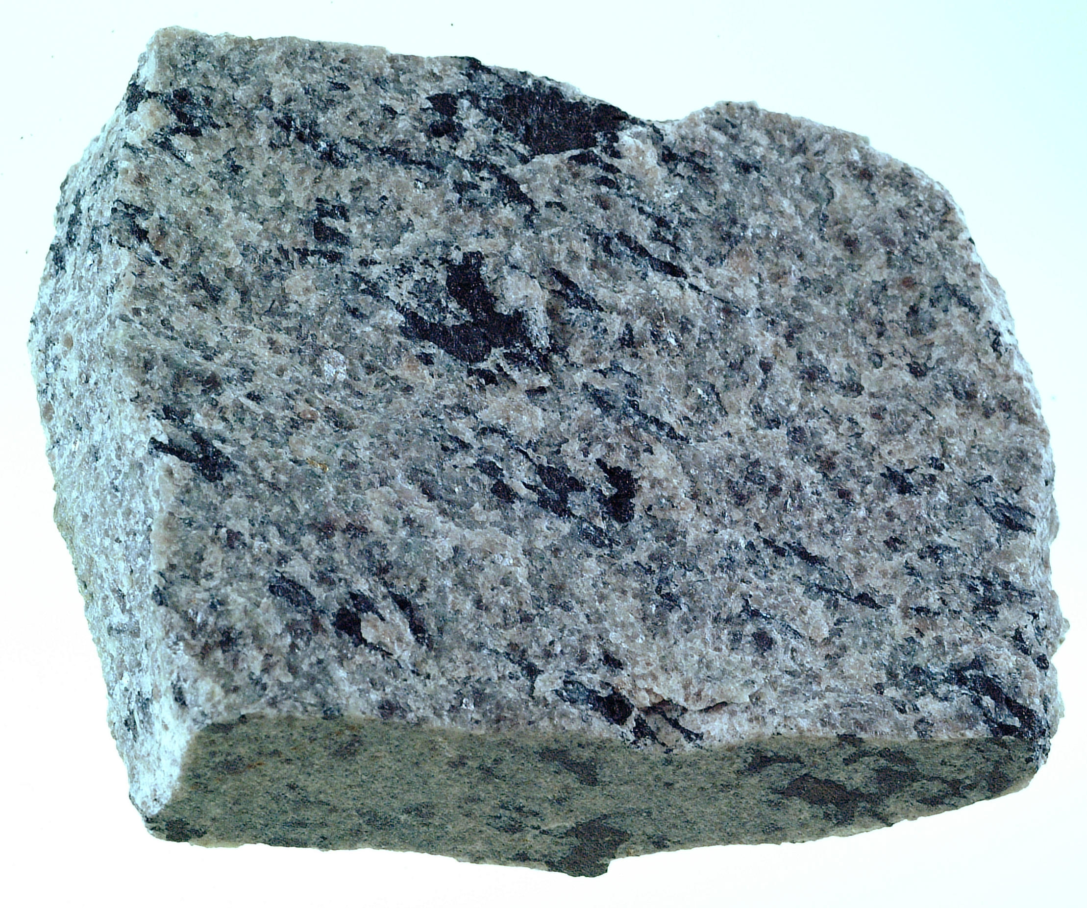
The hand sample in Figure 16.37 is anorthosite from the near Elizabethtown, New York, in the Adirondack Mountains. It is almost entirely an aggregate of plagioclase but contains some black enclaves of clinopyroxene. The Adirondacks are well known for containing one large, and several smaller, anorthosite massifs.
The XP thin-section view in Figure 16.39 above is of a different anorthosite. The view is almost entirely plagioclase, but a few small grains of orthopyroxene (with low 2nd-order interference colors) can be seen near the center of the photo.
▪Norite
|
|
|
|

Norite, a kind of rock dominated by orthopyroxene and plagioclase, is very similar to gabbro. The difference is that norites contain orthopyroxene and gabbros contain clinopyroxene. The close-up hand-sample view above is of a norite from South Africa’s Bushveld Complex. The rock contains black orthopyroxene and light-colored plagioclase that is somewhat stained by hematite. The thin-section views are of a different norite, one from Germany.
In the PP thin-section view, the lath-like pyroxene grains display high relief, good cleavage, and, if you look closely, you can see a hint of the light-pink to green pleochroism that often characterizes orthopyroxene. The clear grains are all plagioclase. Very minor amounts of brown biotite and an opaque mineral are also present.
In crossed polars, the orthopyroxene has up to low 2nd-order interference colors. A few small grains display higher-order colors; these are clinopyroxene grains that are difficult to pick out in the plane-polarized view. The plagioclase grains exhibit typical plagioclase twinning.
▪Norite or Gabbro?
|
|
|
|

| ⇒Link to 3D rotatable sample of the hand sample above |
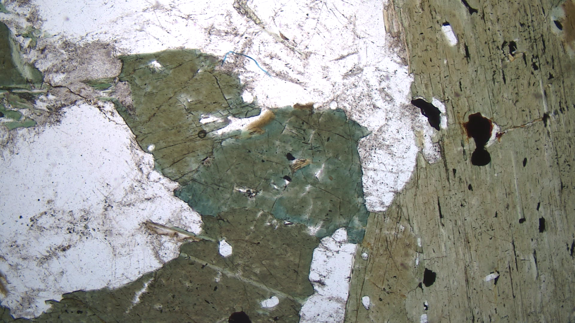
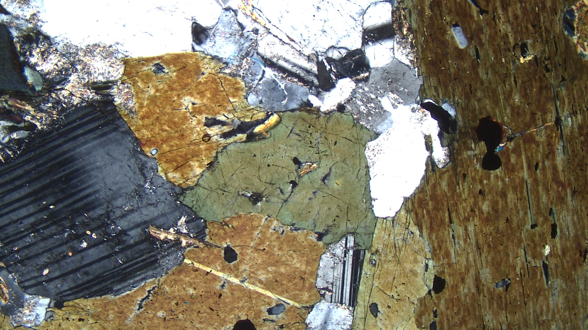
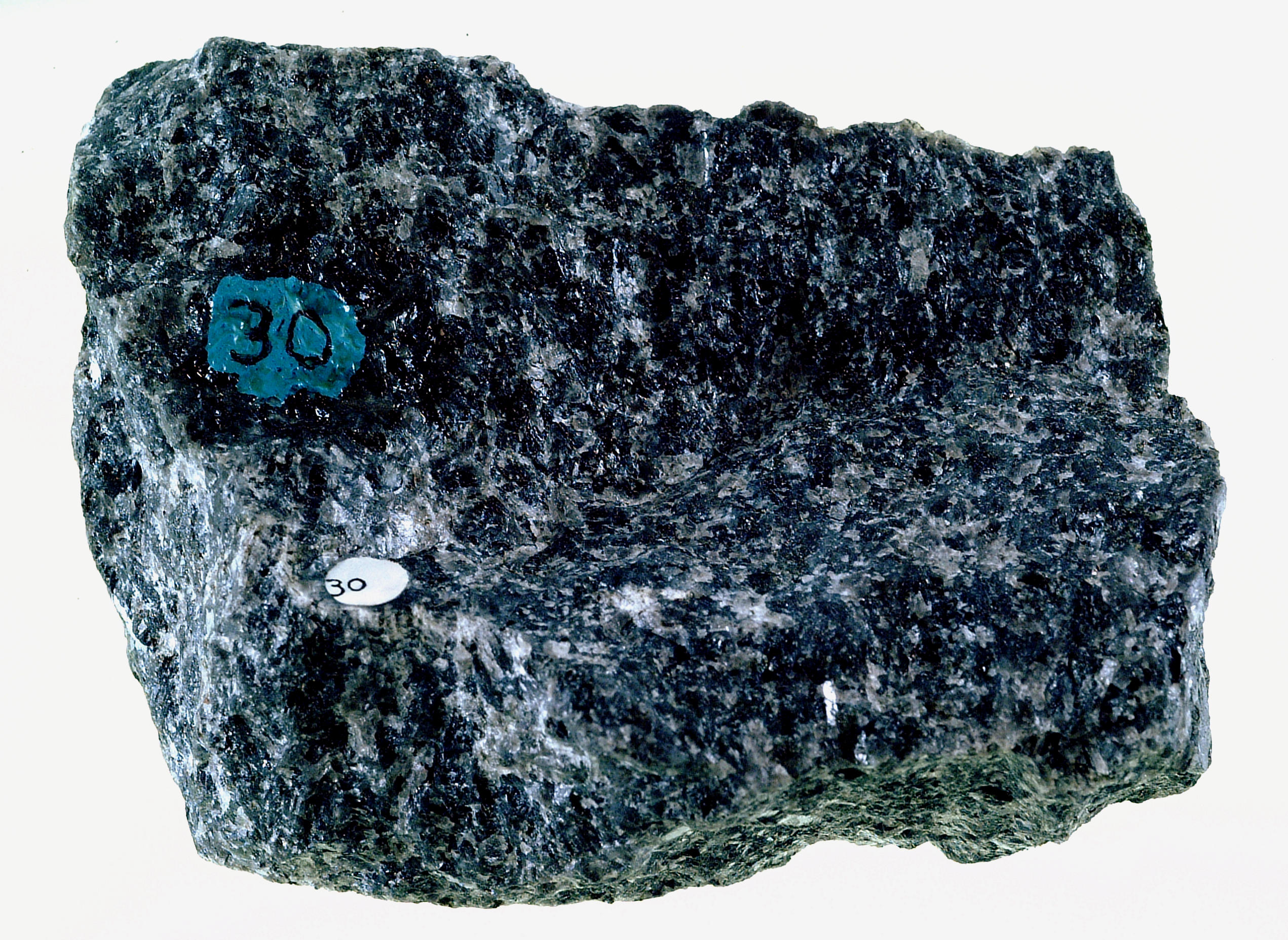
This rock (Figure 16.43) came from Wards Science labeled as a norite from Wollaston Township, Ontario. Maybe it is a norite, but norites contain mostly orthopyroxene and plagioclase. I think this rock may contain clinopyroxene instead of orthopyroxene. In which case, it is another example of gabbro.
The major primary minerals in this specimen are plagioclase and pyroxene. But, the light-green pyroxene is altering to brown biotite and green hornblende. Abundant magnetite is also present. Most, not all, orthopyroxenes display pleochroism from light-green to light-pink. The pyroxenes in this rock do not, and that is one reason I suspect they are clinopyroxene.
In crossed polars, the plagioclase appears zoned and some grains have altered cores. Biotite’s interference colors are up to 2nd order but the brown color of the grains makes interference colors hard to see. The pyroxene has up to 3rd-order interference colors, which would be unusual if it was orthopyroxene. That is the main reason that I suspect that the pyroxenes are augite and that Wards has misidentified the sample. On the other hand, elongate pyroxene grains have near parallel extinction, and parallel extinction is commonly one way to distinguish orthopyroxene from clinopyroxene.
▪Diabase
Diabase is a mafic rock with composition equivalent to gabbro and basalt. But diabase is coarser grained than most basalt and finer grained than most gabbro. The dominant minerals in the diabase shown below, like all diabase, are light-colored plagioclase and dark-colored clinopyroxene. Both can be seen in the hand sample.
|
|
|
|
| ⇒Link to 3D rotatable sample of this diabase |
More than half of the thin-section view above shows plagioclase. It is clear in plane-polarized light and displays classic twinning in crossed polars. Augite appears as gray high-relief grains in the PP view; one long augite lath runs diagonally across the right part of the photo. The augite has altered to brown and green biotite on some edges. One large magnetite grain is present on the right side of the photo.
The pyroxene has up to high 2nd-order interference colors (XP view). The large pyroxene lath in the center of the view is twinned. The upper half of the grain also contains thin parallel exsolution lamellae giving a poorly formed “herringbone structure.”

The rock seen in outcrop in this photo is a diabase that is part of the Palisades Sill in New Jersey and New York. Figure 16.49 is an outcrop of the same rock. This sill forms cliffs for 65 km along the Hudson River and is 300 m thick. Figure 7.28 shows another photo of the sill.
▪Troctolite
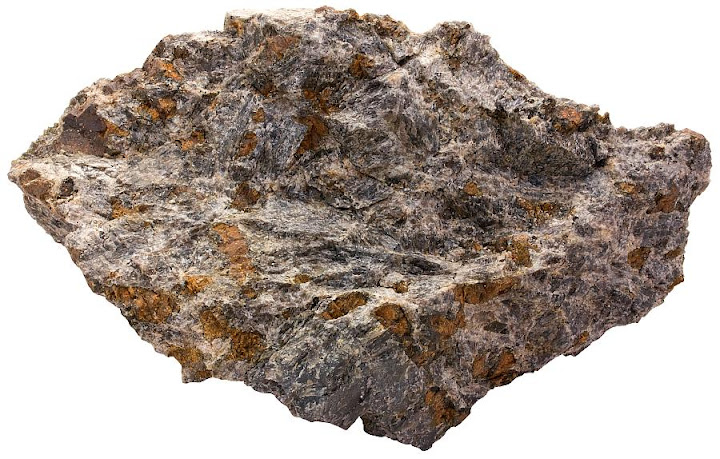
Troctolites are rare rocks that consist primarily of olivine and plagioclase. Minor amounts of pyroxene may also be present. The hand sample seen below comes from the Lofoten Islands in western Norway. Olivine is quite unstable at Earth’s surface and weathers quickly. So, most troctolites, like this one, contain olivine that is not olivine’s typical green color. In this sample, the original olivine crystals appear brown due to staining by hematite.
|
|
|
|
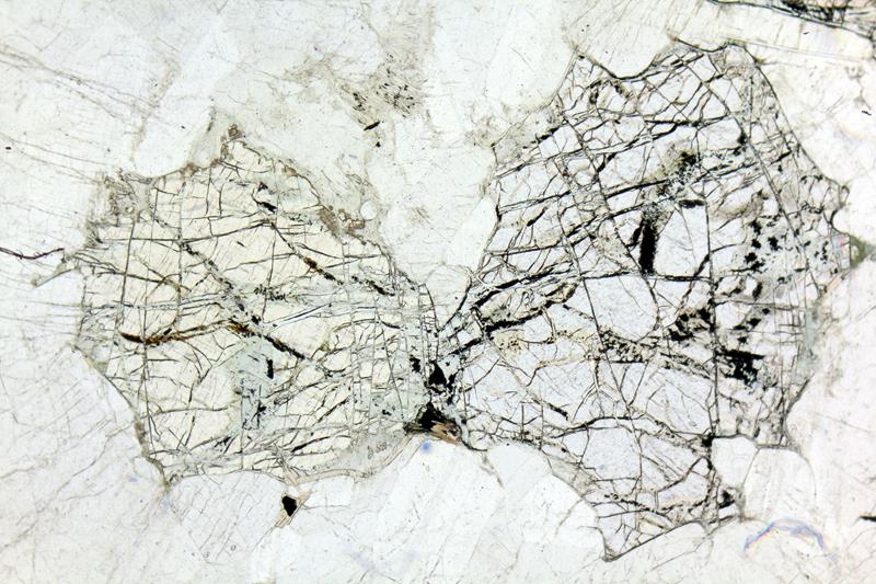
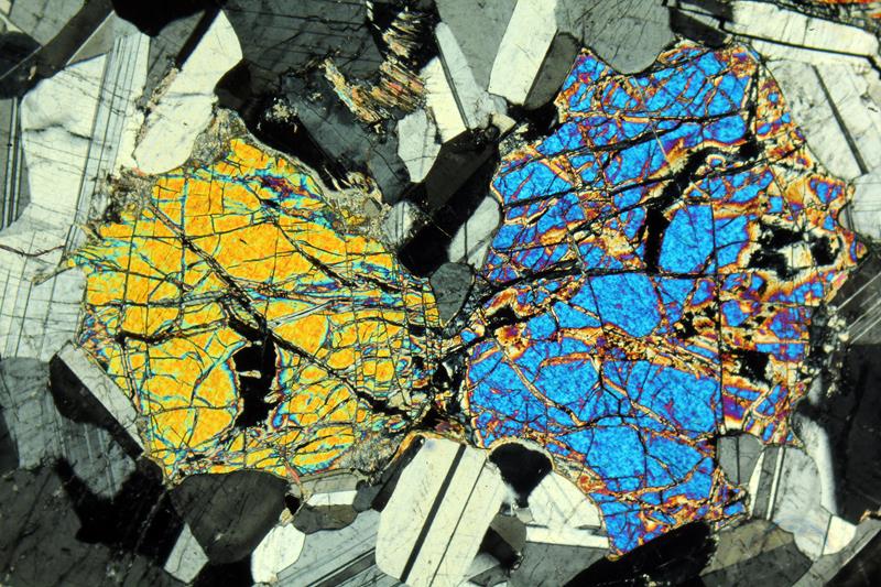
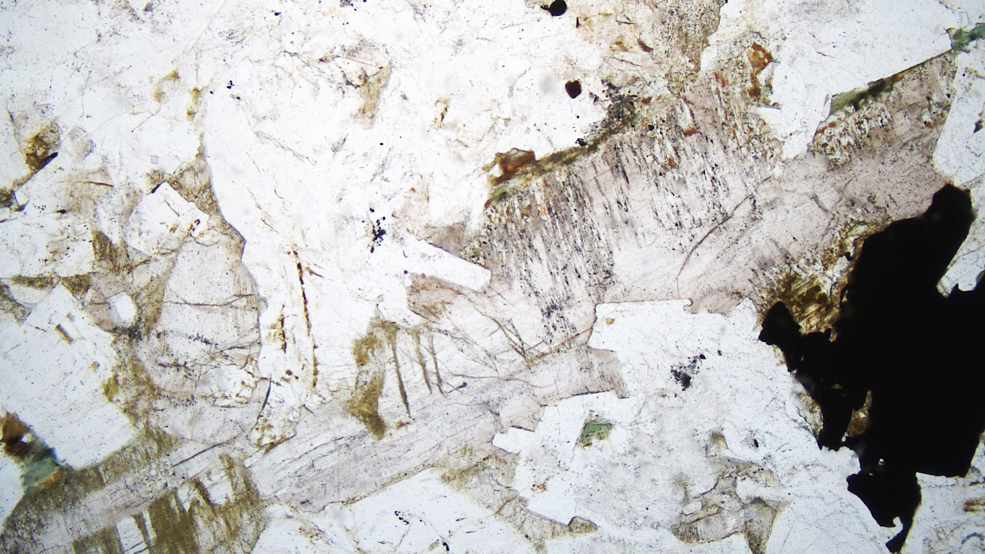
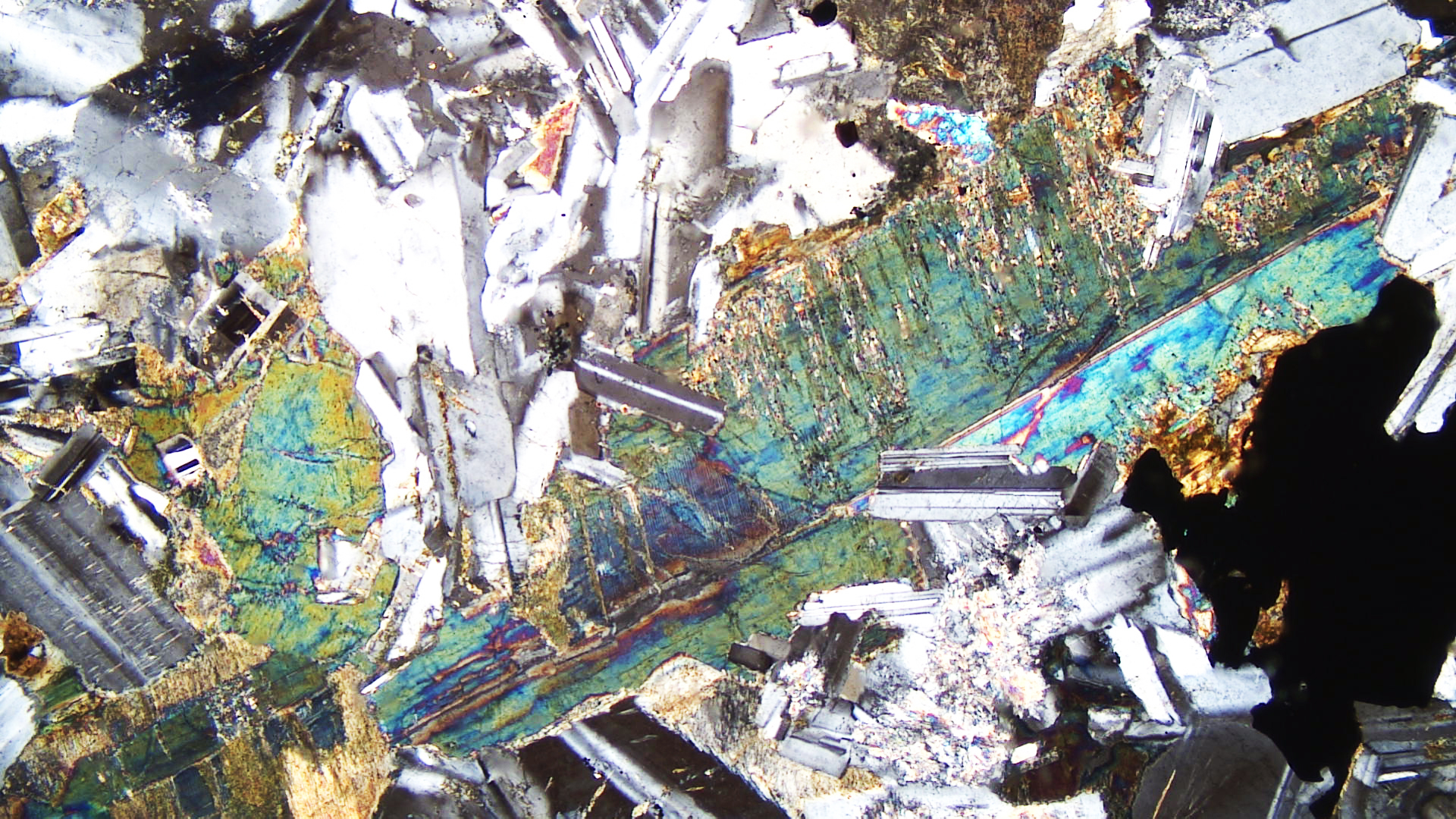
The two thin-section views above show a troctolite containing two large fractured olivine grains. The clear mineral around them is plagioclase. In crossed polars, the olivine has strong 3rd-order colors and the plagioclase has 1st-order colors and displays well-developed twinning.
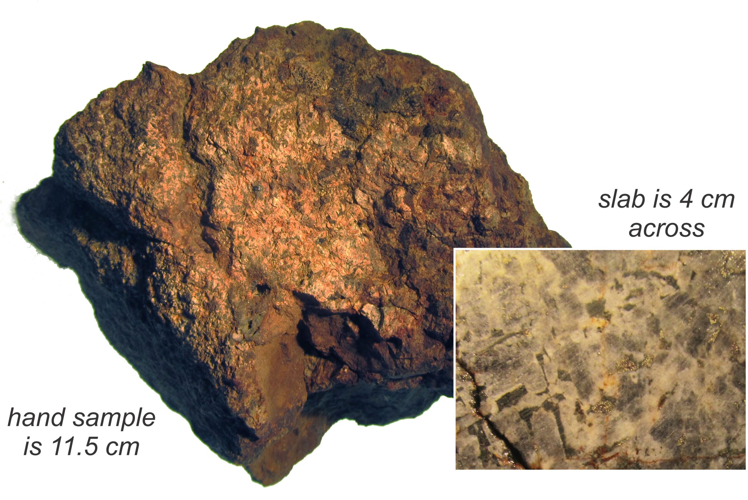
The image seen on the right is a troctolite from the western margin of Minnesota’s Duluth Complex. This troctolite hosts a sulfide-rich ore deposit that has attracted the attention of mining companies, but also of environmentalists who do not want mining to take place near the Boundary Waters Wilderness. The rock contains abundant sulfides and its weathered surfaces are altered to a rusty red color. The inset photo shows a polished slab of the rock’s interior. The interior appears relatively fresh compared with the exterior. Plagioclase is white and olivine is dark gray. Some olivine crystals have a vague orthorhombic shape.
|
|
|
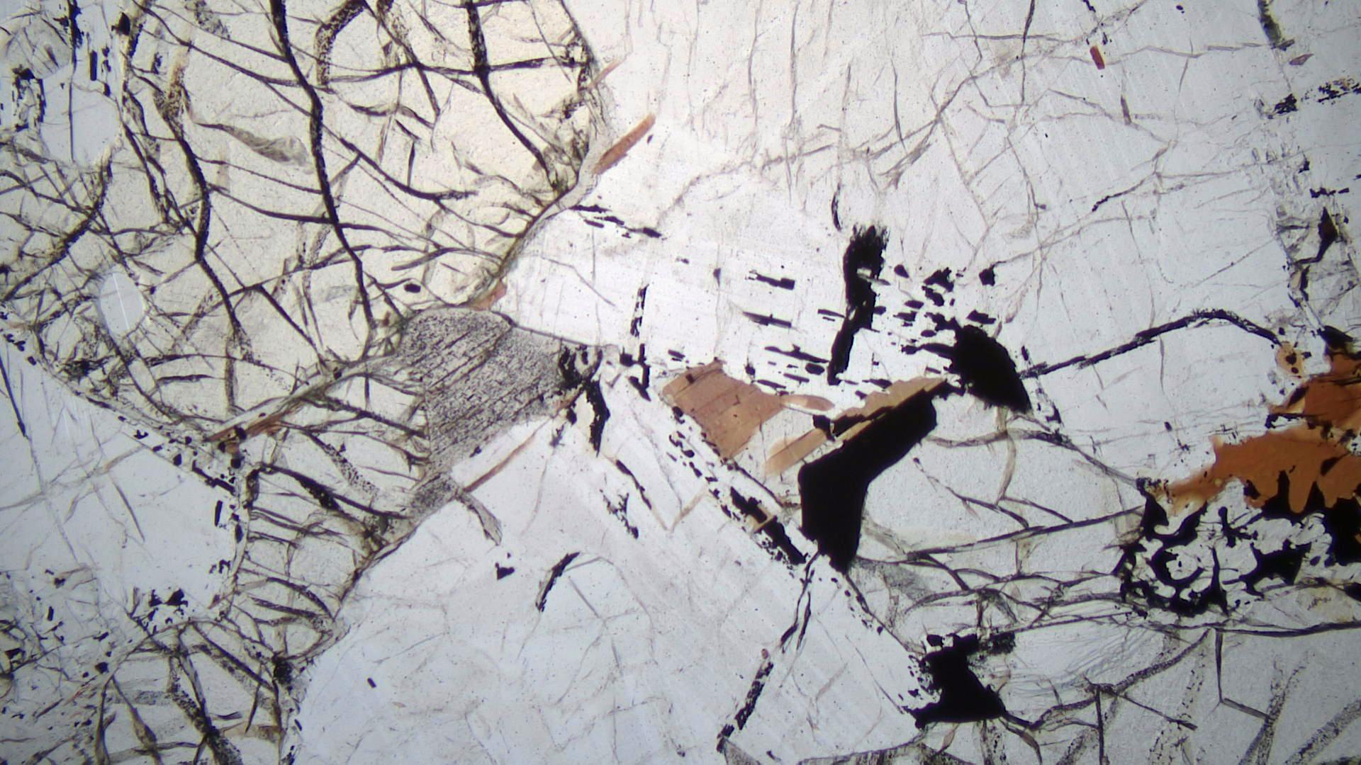
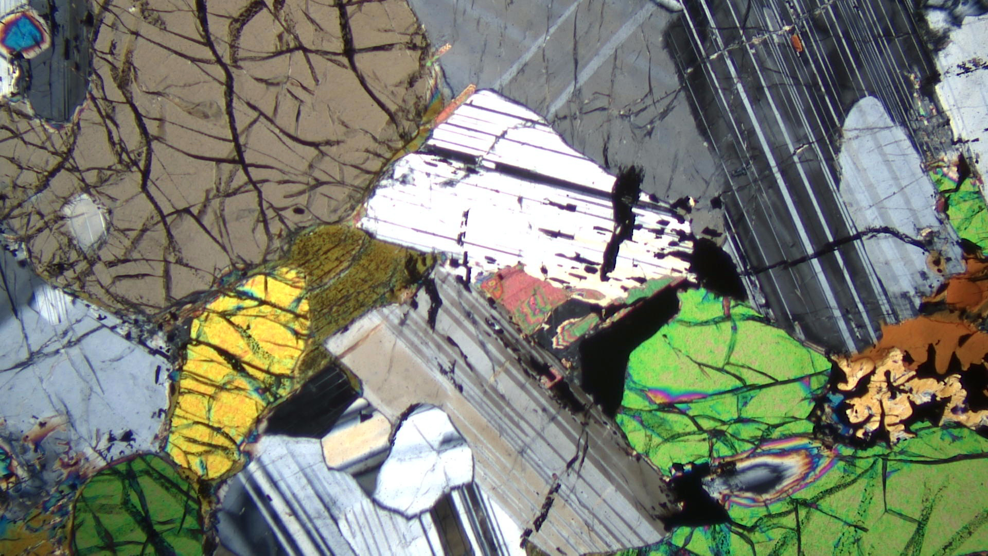
The thin-section views seen here are of the same Duluth Complex troctolite in the photo above. The plane-polarized view contains mostly high-relief olivine with many fractures, and clear plagioclase. Minor amounts of brown biotite and some opaque sulfide grains are also evident. A single grain of gray clinopyroxene is just left of the center of the photo, adjacent to olivine. In crossed polars, olivine has up to 3rd-order interference colors, twinned plagioclase shows first order colors, and the clinopyroxene appears near to extinction.
16.1.4 Ultramafic Plutonic Rocks
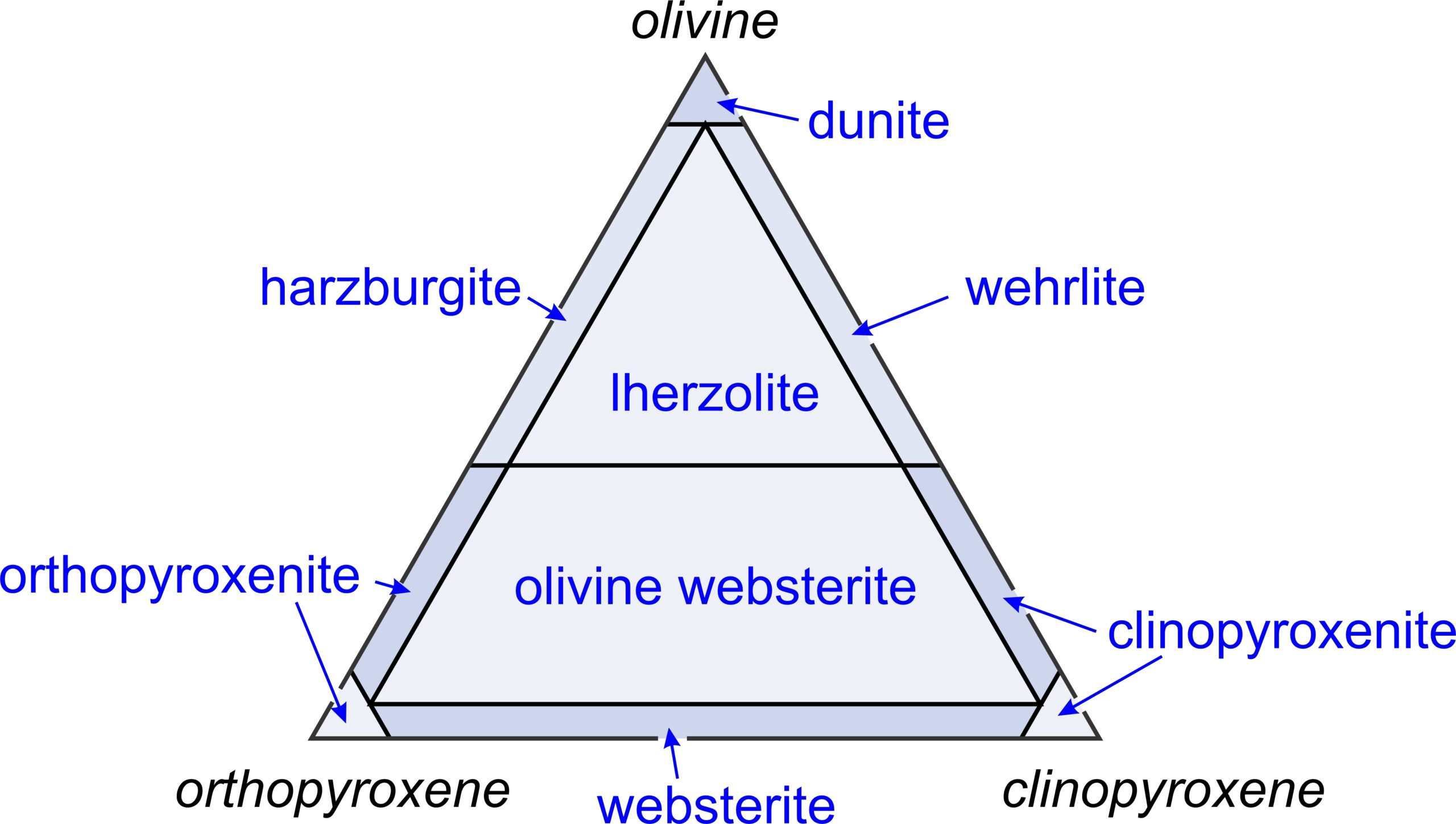
16.56 IUGS classification system for the most common kinds of ultramafic rocks
Figure 16.56 is the IUGS classification system for different kinds of ultramafic rocks. Ultramafic rocks contain some combination of olivine, orthopyroxene, and clinopyroxene. Rocks that contain more than 40% olivine (and plot in the top half of the IUGS triangle) are peridotites, including harzburgites, lherzolites, wehrlites, and dunites. Pyroxenite is the generic name for ultramafic rocks that contain less olivine, including
orthopyroxenites, clinopyroxenites, and websterites.
▪Dunite
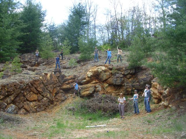
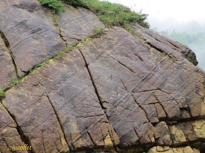
Ultramafic rocks of various sorts, including dunite and websterite, are found in the Balsam to Spruce Pine region of the Blue Ridge Mountains east of Smoky Mountain National Park. (Websterites are named after the town of Webster, just a few kilometers from Balsam.) These ultramafic and associate rocks are pieces of the lower crust and mantle that became uplifted and part of North America during mountain building. Figure 16.60 is a photo of a dunite outcrop from that region. The dunite in the photos below comes from the same area.
|
|
|
|
| ⇒Link to 3D rotatable image of the dunite shown above |
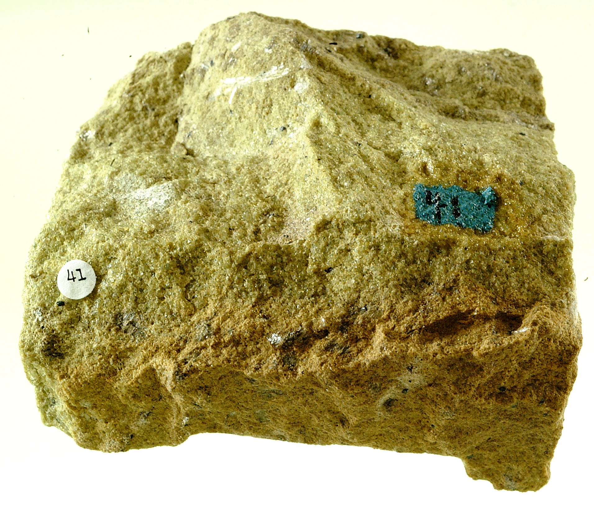
Dunites are ultramafic rocks that, by definition, contain more than 90% olivine. Common accessory minerals include clinopyroxene, orthopyroxene, spinel, ilmenite, magnetite, and chromite. The fine granular rock seen in the photos above is just about entirely made of anhedral olivine crystals. Besides olivine, a few small black grains of chromite are present.
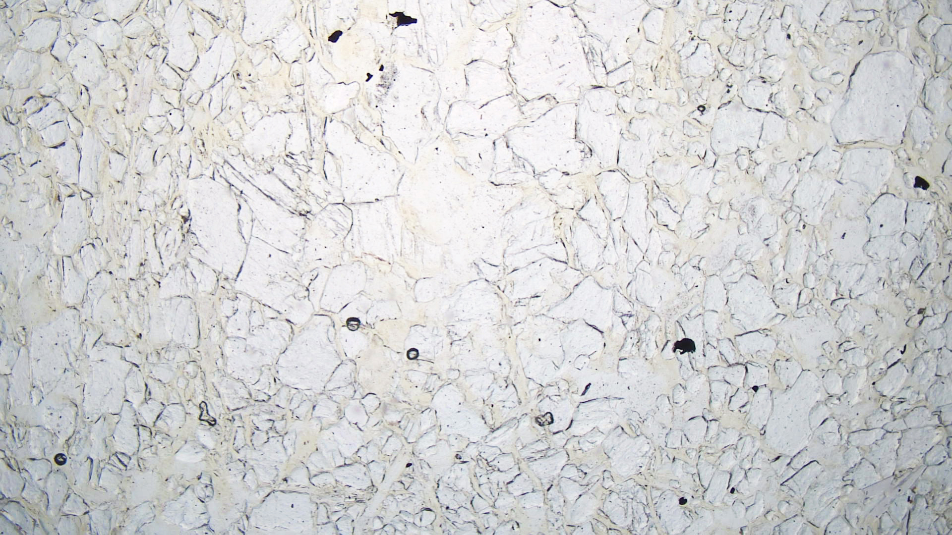
In thin section (PP view) above, olivine appears clear with (maybe) a hint of green. Crystals form a mosaic texture. Chromite appears as small opaque grains in the plane-polarized view. In crossed polars, olivine displays up to the normal (blotchy) 3rd-order colors.
▪Spinel Lherzolite
|
|
|
|

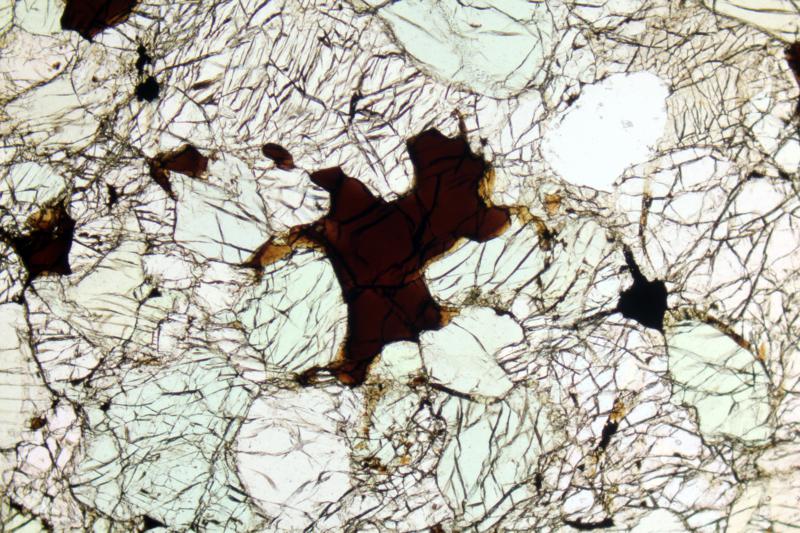
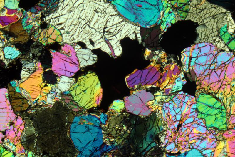
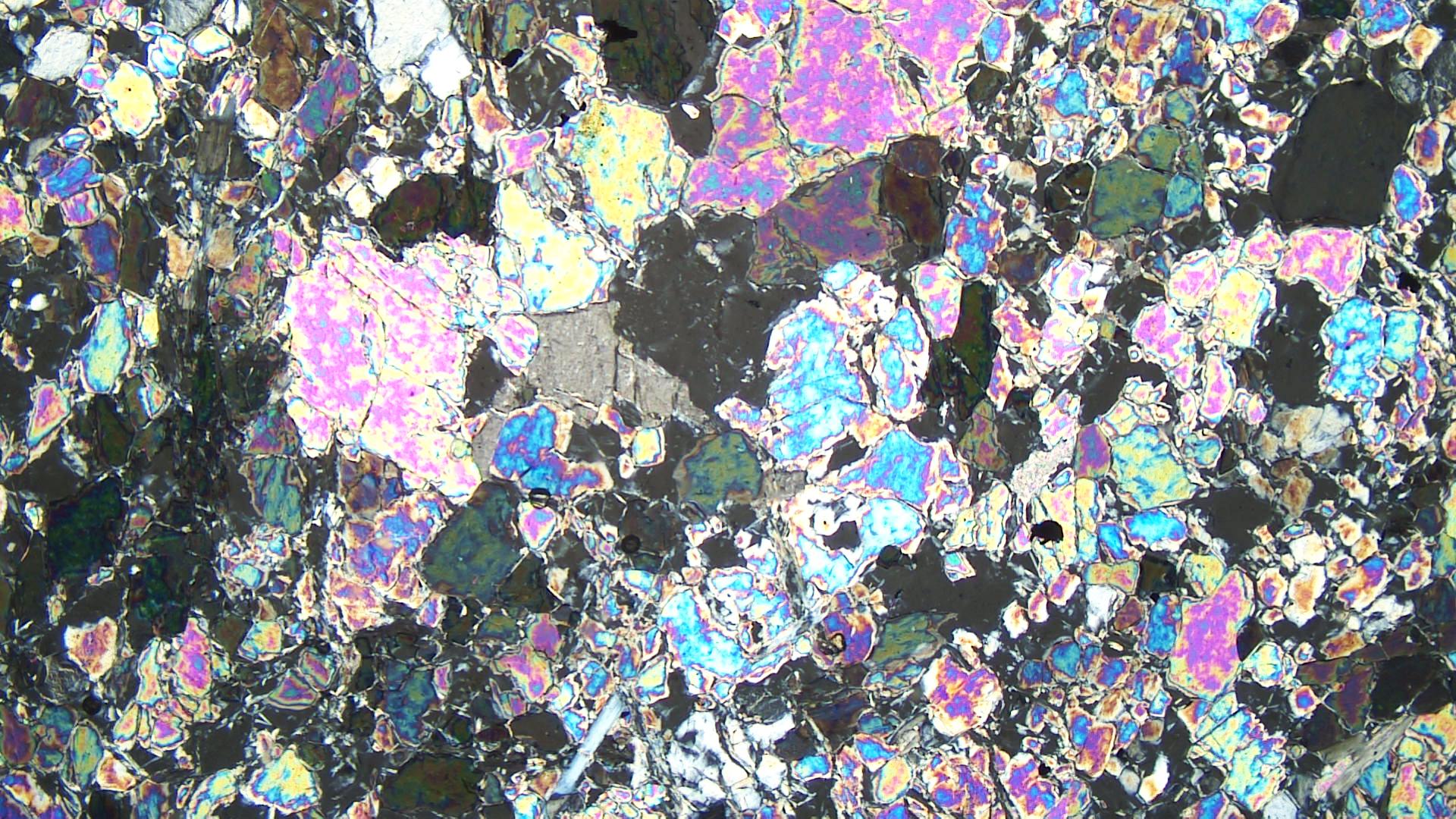
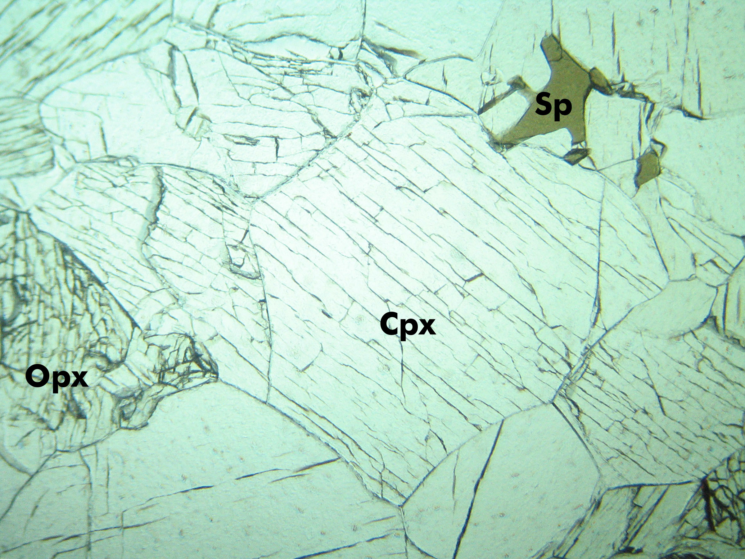
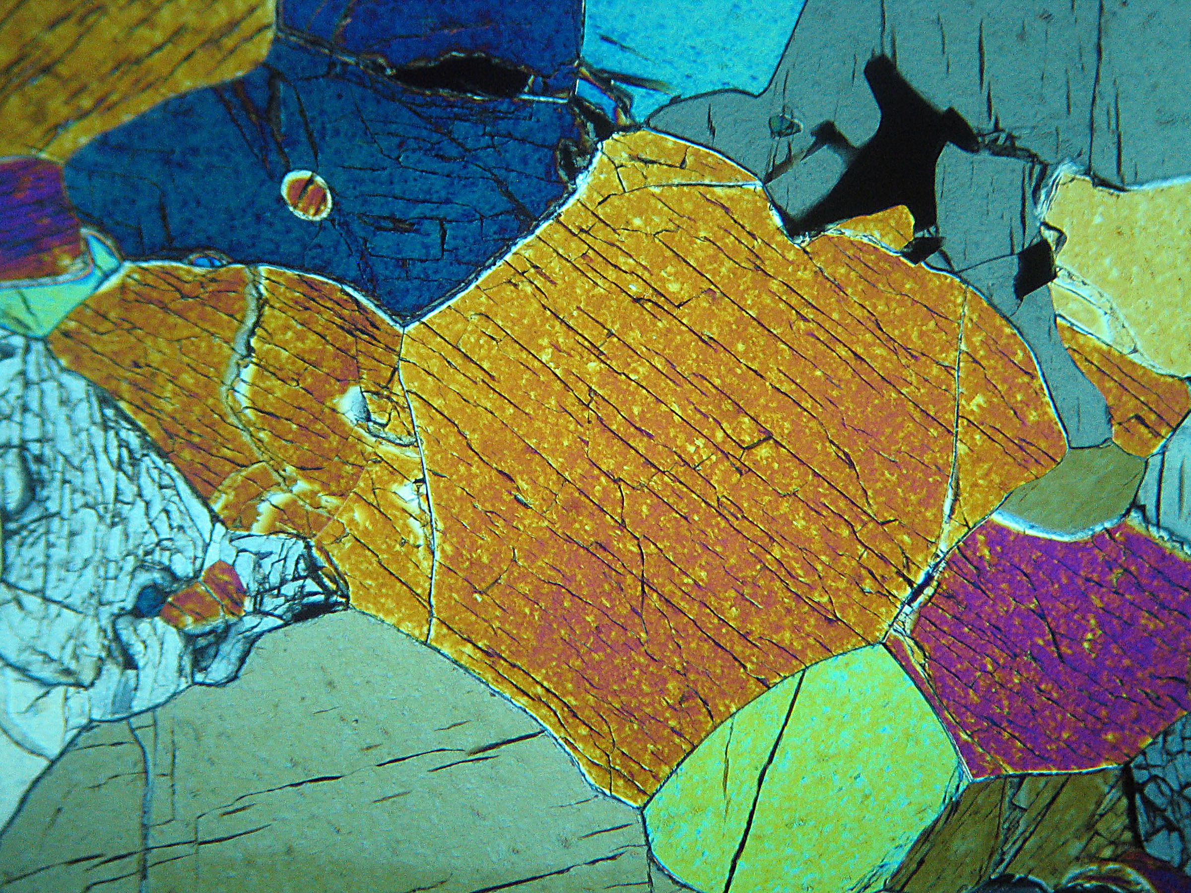
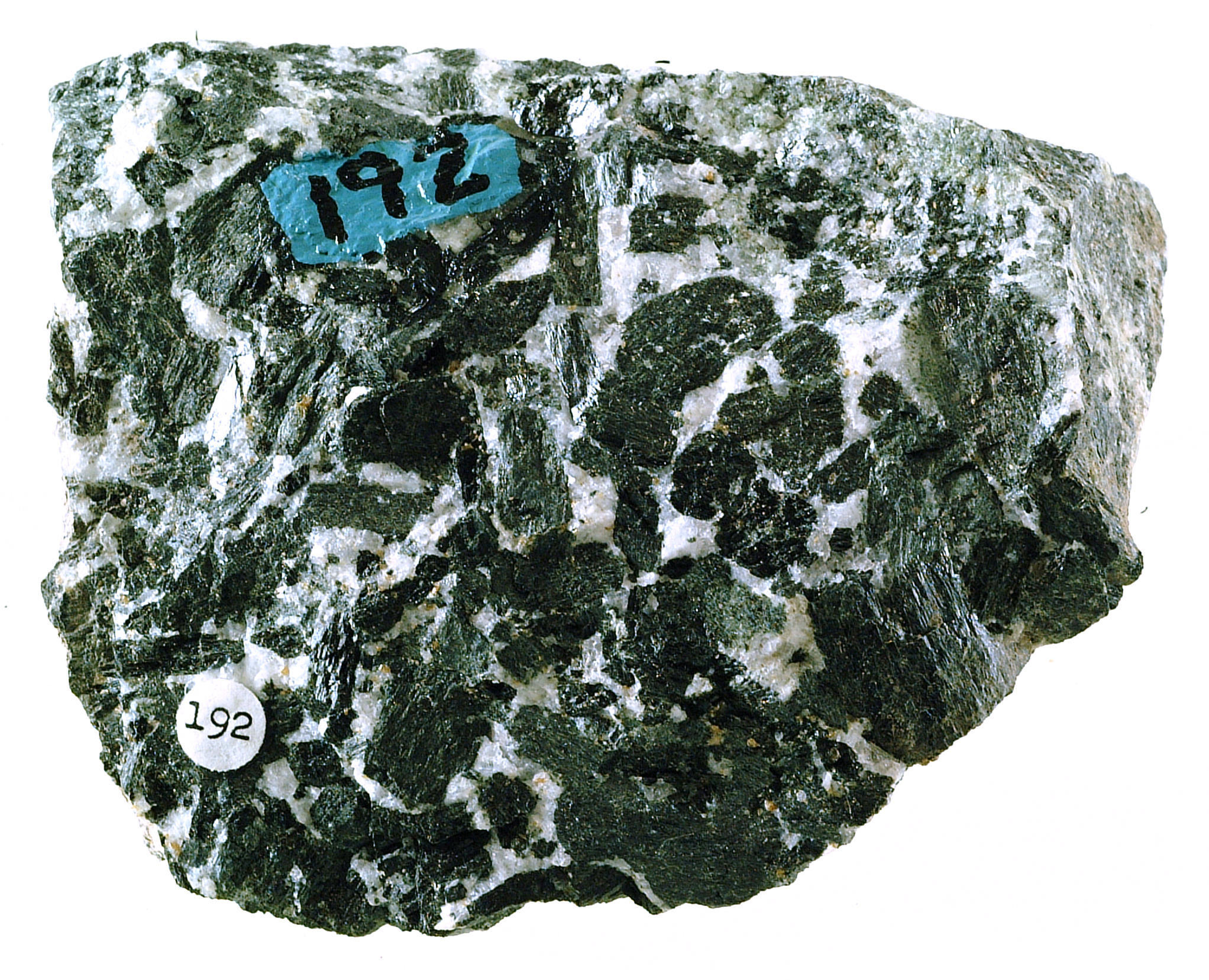
Lherzolites are ultramafic rocks that contain subequal amounts of olivine, clinopyroxene, and orthopyroxene. The hand-sample photo above is a spinel-bearing lherzolite xenolith from Mt. Leura, near Melbourne, Australia. The green ultramafic xenolith is encased in black basalt. Enlarge the photo and you can see much pale green olivine and darker brown/black grains that are orthopyroxene. Clinopyroxene in this rock has a deeper, more emerald-like green color than the olivine but is difficult to distinguish. The minor spinel crystals are gray/brown but cannot be seen in this photo. Figure 16.64 is an outcrop of a different lherzolite, this one in the Lherz Massif of the French Pyroxenes. The thin-section photos above are a spinel lherzolite from that type locality.
The PP view above (Figure 16.62) shows pale-green orthopyroxene, colorless olivine and clinopyroxene, and brown spinel. The olivine grains are blockier than the pyroxene grains, more fractured, and lack cleavage. In crossed polars (Figure 16.63), olivine displays up to 3rd-order interference colors (compared with the 2nd-order colors of the pyroxene). Spinel is isotropic. One large hole in the thin section appears isotropic, too.
▪Wehrlite
|
|
|
|
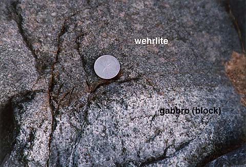
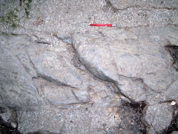
The photo on the left above shows an outcrop that contains gabbro in its bottom half with wehrlite above. Gabbros are dominated by essential plagioclase and clinopyroxene and wehrlites by essential olivine and clinopyroxene. Consequently, wehrlites have a darker color.
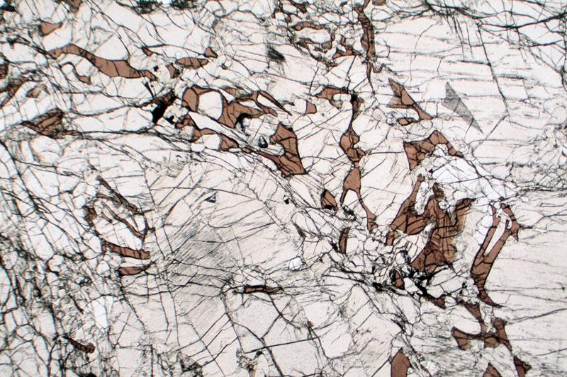
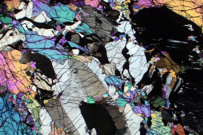
The thin-section views show spinel, clinopyroxene, and olivine in a different wehrlite. The spinel is brown in the PP view; the other minerals are nearly clear and the olivine is hard to distinguish from the clinopyroxene. In crossed polars, pyroxene has lower-order interference colors than olivine. Most of the pyroxene grains display only 1st-order colors. One large pyroxene grain (just left of center in the bottom half of the thin-section views) displays 1st-order gray interference colors, twinning, and exsolution in the XP view, giving it a partially developed herringbone texture. The more birefringent olivine grains contain more fractures and more variable/patchy interference colors (up to 3rd order) than the pyroxene grains. The spinel is isotropic.
▪Clinopyroxenite and Websterite
|
|
|
|
The hand specimen seen above in Figure 16.68 is a clinopyroxenite from the Serra Negra-Salitre Complex in southeastern Brazil. This rock is almost entirely clinopyroxene, but some pyroxenites contain both clinopyroxene and orthopyroxene. Websterite, a variety of pyroxenite, is an ultramafic rock that contains subequal amounts of both pyroxenes. Spinel, plagioclase, and other accessory minerals may be present in pyroxenites but are generally minor.
The thin-section views are photos of a websterite from Kilbourne Hole, New Mexico (discussed in Chapter 5). The rock is more than half clinopyroxene; the remainder is mostly orthopyroxene. The two pyroxenes appear similar in the PP view. The single large orthopyroxene grain, however, has slightly greater relief and is oriented to show more cleavage. In crossed polars, the clinopyroxene displays much higher interference colors (up to upper 2nd order) than the orthopyroxene (which shows its typical 1st-order colors). Minor spinel (green in the PP view) is present and isotropic in the XP view.
16.1.5 Feldspathoid-Bearing Plutonic Rocks
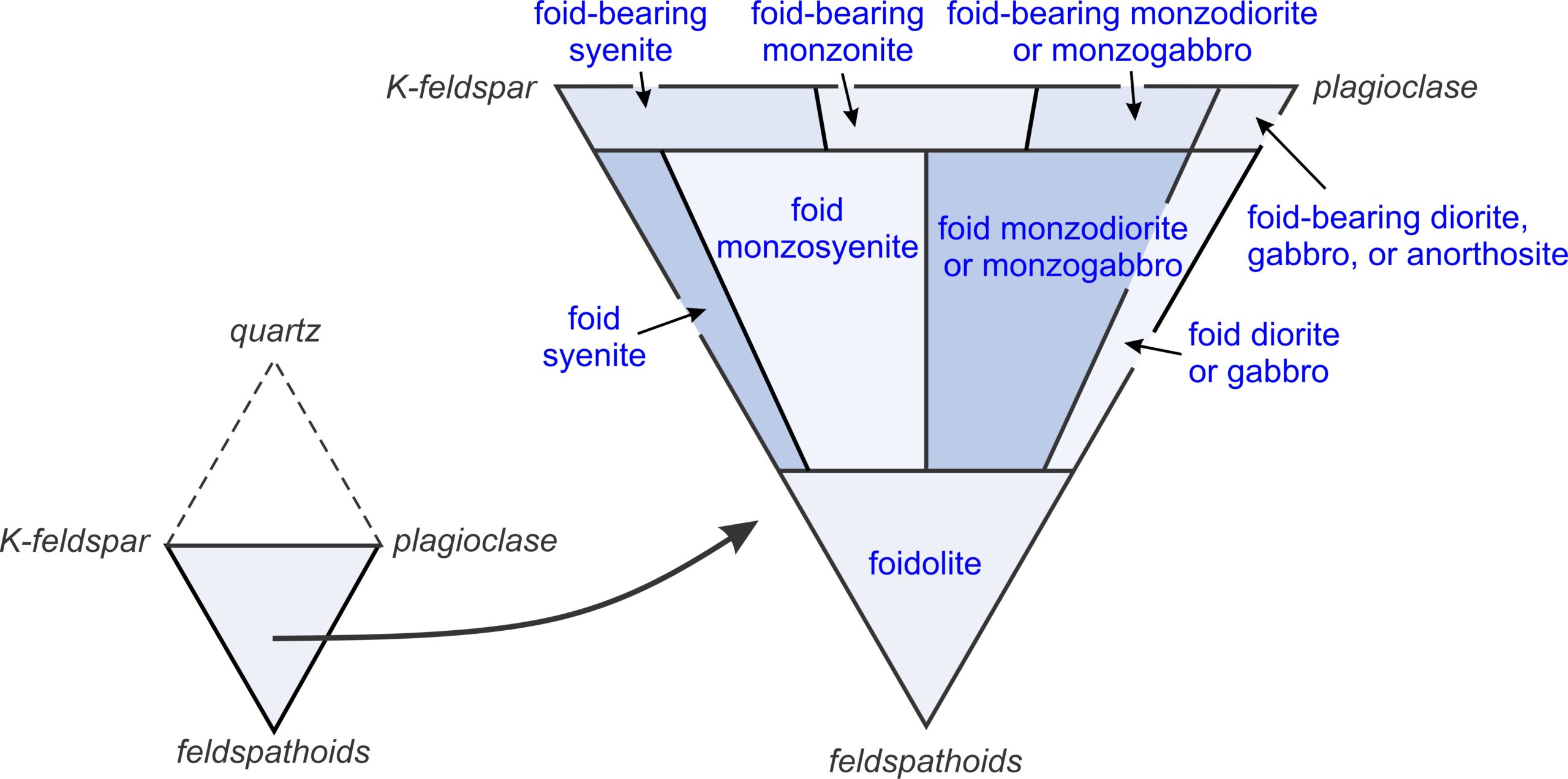
Igneous rocks with relatively low silica contents may contain feldspathoids, such as nepheline or leucite. These minerals resemble feldspars in composition and character but contain less silica. Feldspathoidal rocks do not contain quartz. The IUGS classification system (a slightly simplified version is in Figure 16.71) names these rocks based on the relative amounts of K-feldspar, plagioclase, and feldspathoid they contain. For specific rock names, we replace the term foid in Figure 16.71 with the name of the feldspathoid present. Most commonly, the feldspathoid is leucite, nepheline, or sometimes sodalite.
Feldspathoid-bearing rocks are uncommon, and most contain significantly more feldspar than feldspathoids. The exceptions are mostly rare kinds of volcanic rocks.
▪Nepheline Diorite
|
|
|
|
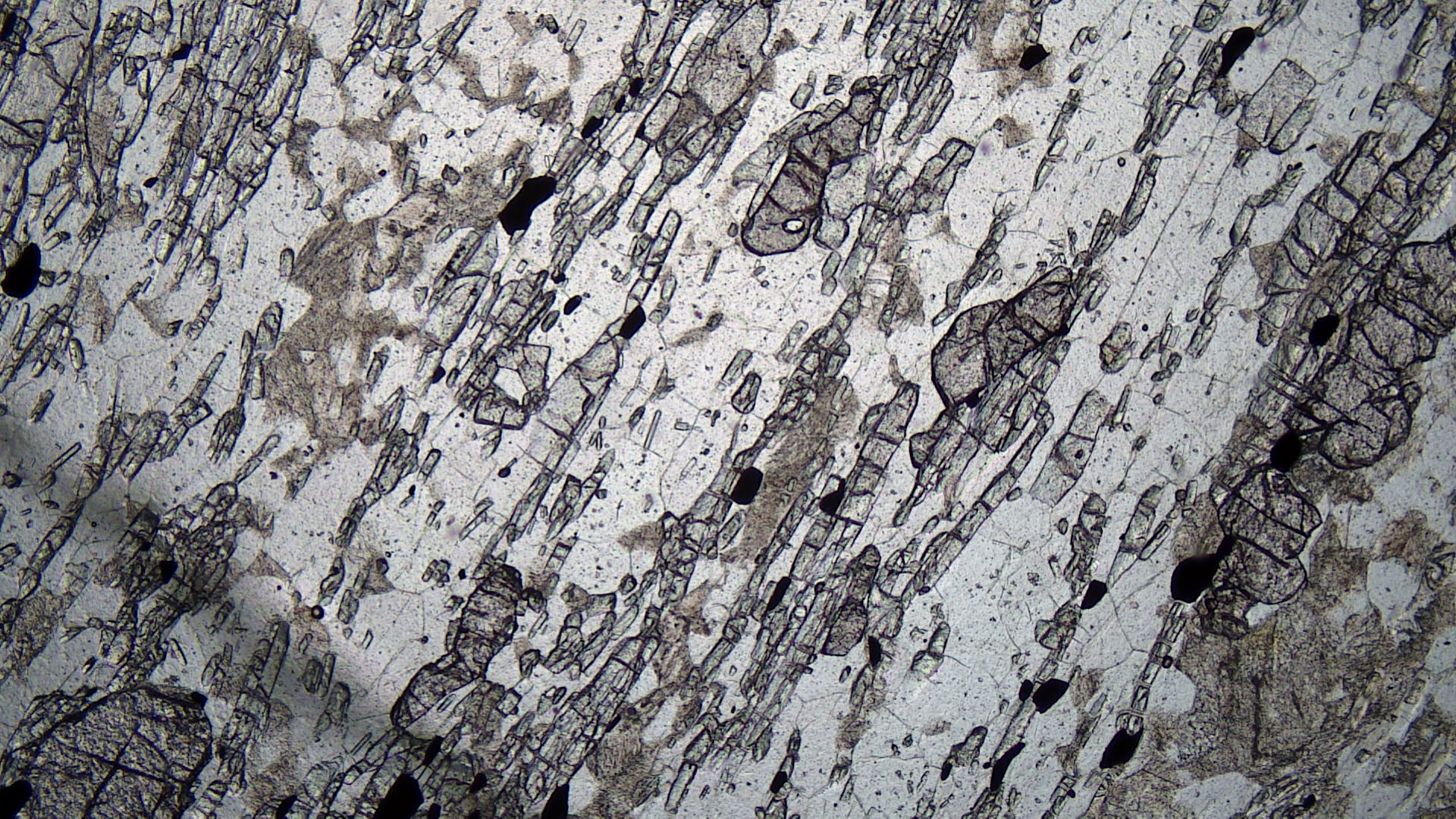
The outcrop photo above shows a horizontal nepheline syenite dike that has intruded nepheline diorite (coarser-grained rock above and below the dike). This outcrop is in Monteregian Hills near Quebec City.
The thin-section views above are photos of a nepheline diorite from Russia’s Kola Peninsula. The blocky nepheline crystals contain many inclusions and holes. They are nearly clear in the PP photo and, because nepheline has very low birefringence, are light- to dark-gray in the XP view. Fine blue-green clinopyroxene (aegirine) and plagioclase make up the groundmass. A few of the pyroxene crystals, especially some near the top-right corner of the photos, are large enough to show 2nd-order interference colors.
▪Nepheline Syenite
The main minerals in nepheline syenites are nepheline and alkali feldspar. Mafic minerals, including clinopyroxene, amphibole, and biotite are typically present as accessories. The photos below show a nepheline syenite from Bancroft, Ontario. The rock contains subequal amounts of nepheline and albite. Mafic minerals include biotite and hastingsite (a calcic hornblende). The rock also contains calcite.
|
|
|
|
| ⇒Link to 3D rotatable sample of this syenite |
Two prominent, somewhat blocky, opaque magnetite grains appear in the thin-section views. In PP light, nepheline, albite, and calcite appear nearly clear. Several large calcite crystals, however, have higher relief than the other minerals. Three needles of dark-brown colored hastingsite stand out. In crossed polars, calcite has very high-order interference colors, some twinning, and a pebbly texture. Other grains displaying high-order interference colors are cancrinite, a rare carbonate-silicate mineral. Albite contains prominent lamellar twins and the nepheline is blocky and displays good cleavage.
▪Nepheline Syenite
The hand specimen in the photo below is a different kind of nepheline syenite compared with the one described above. This specimen, from Montana’s Crazy Mountains, contains abundant clinopyroxene. The thin-section views are of the same rock. Much of this rock is made of euhedral to subhedral pyroxene crystals. Nepheline and sanidine make up most of the rest. Very minor brown biotite, green hornblende, and magnetite are also present.
|
|
|
|

| ⇒Link to 3D rotatable sample of this nepheline syenite |
In plane-polarize light, the pyroxene appear as well-formed, almost clear, elongate crystals. In places it is altering to a green amphibole. In crossed polars, the pyroxene displays 2nd-order colors, and a few grains show hints of twinning. The nepheline and sanidine – hard to tell apart – are clear in the PP view and display 1st-order interference colors in the XP view.
▪Sodalite Syenite
|
|
|
|
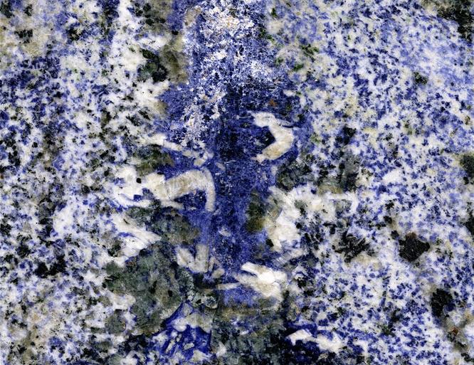
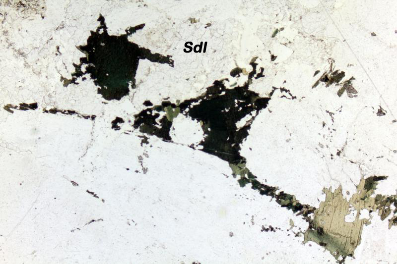
The rock slab seen in the photo on the left above is a popular stone used for counter tops and other purposes. The rock comes from Bahia State, Brazil. Its commercial name is Azul Bahia Granite, but it is actually a syenite that contains sodalite. The large visible crystals are blue sodalite, white to gray K-feldspar, and leucite. Sodalite is one of the few feldspathoids that have a distinctive color.
Figures 16.82 and 16.83 are thin-section photos of a different sodalite syenite – one from Greenland. Sodalite is clear in PP light but is isotropic, so it appears black in the XP view. The other minerals present are biotite (brown and greenish to almost black in the PP view) and clear alkali feldspar. The biotite has weak 2nd-order interference colors that are almost masked by the color of the mineral. The alkali feldspar has its typical 1st-order colors.
▪Leucitite
|
|
|
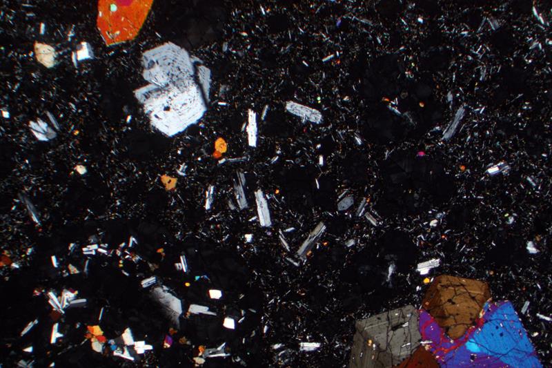
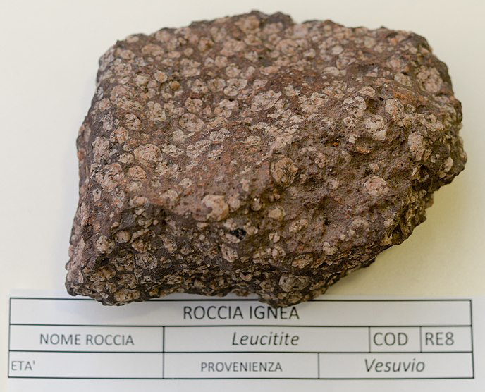
Leucitites are rocks that contain more than half leucite, generally with clinopyroxene, and no, or small amounts of feldspars. These rocks are exceptionally rare, and most are volcanic or hypabyssal (shallow level intrusions).
The thin-section photos in Figures 16.84 and 16.85 show a leucitite. The PP view contains clear leucite and gray-green high-relief clinopyroxene. Leucite has very low birefringence and has black to dark-gray interference colors in the XP view. The clinopyroxene shows up to high 2nd-order colors.
▪Nephelinite
|
|
|
Nephelenites are rocks that contain abundant nepheline and small amounts of feldspars. Like leucitites, these are rare rocks, and most are volcanic or hypabyssal. Figures 16.86 and 16.87 are photos of a nephelinite in thin section. It contains clear nepheline, brown augite crystals, and an opaque mineral. In the XP view, nepheline’s interference colors are up to 1st-order gray; the augite shows 2nd-order colors.
16.2 Volcanic Rocks
Some effusive rocks contain quartz, alkali feldspar, and plagioclase, singly or in combination, and minor or no glass. If we can identify these minerals and their relative proportions, the proportions yield an IUGS rock name using the QAPF (quartz-alkali feldspar-plagioclase-feldspathoid) diagram on the left in Figure 16.88.
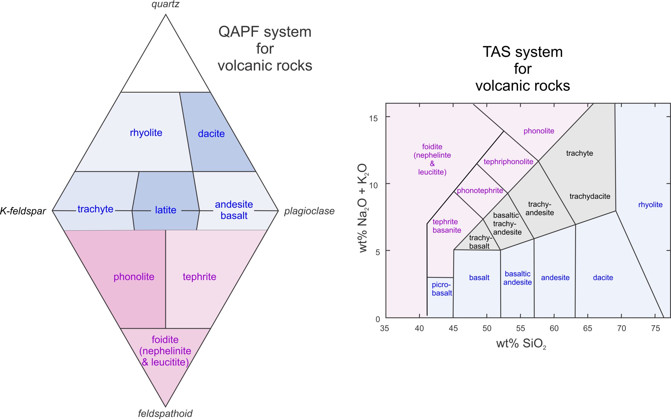
However, many volcanic rocks contain glass in lieu of minerals, and often minerals, if present, are so fine-grained that they are difficult to identify. So, the IUGS has developed a different naming scheme that assigns names to rocks based on their chemical composition (diagram on the right in Figure 16.88). We discussed these two systems more fully in Chapter 4.
Most volcanic rocks have compositions that plot in the blue regions on the two diagrams above. Those that are relatively silica-poor and alkali-rich contain feldspathoids and plot in the violet regions. The two systems yield similar rock names for most volcanic rocks. The exceptions are rocks with compositions that plot in the gray region in the TAS diagram above. And, in the absence of a chemical analysis, assigning names can be problematic. It is not, however, fruitful to go into more detail in this chapter.
16.2.1 SiO2-Rich Volcanic Rocks
▪Rhyolite Tuff

Rhyolite is a kind of silicic volcanic rock with a composition equivalent to granite. Some rhyolite crystallizes from lava and may contain phenocrysts of quartz and alkali feldspar, sometimes with lesser amounts of plagioclase and biotite phenocrysts. The phenocrysts are surrounded by a very fine-grained or glassy groundmass. More commonly, rhyolitic rocks are tuffs, like the tuff seen in Figure 16.89, that form by compaction and cementation of volcanic ash and other pyroclastic debris produced during an explosive eruption. These tuffs may be largely or entirely made of glass.
|
|
|
|
| ⇒Link to 3D rotatable image of this rhyolite |
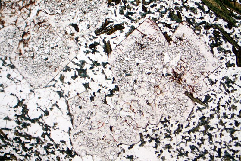
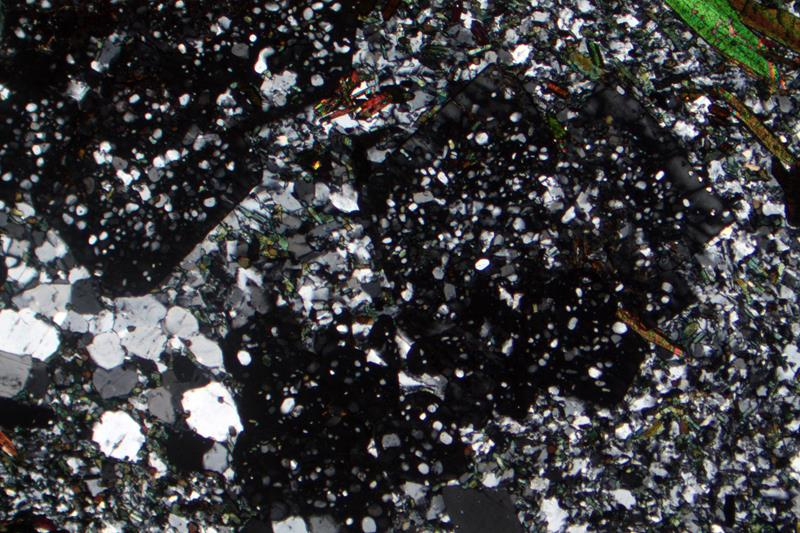
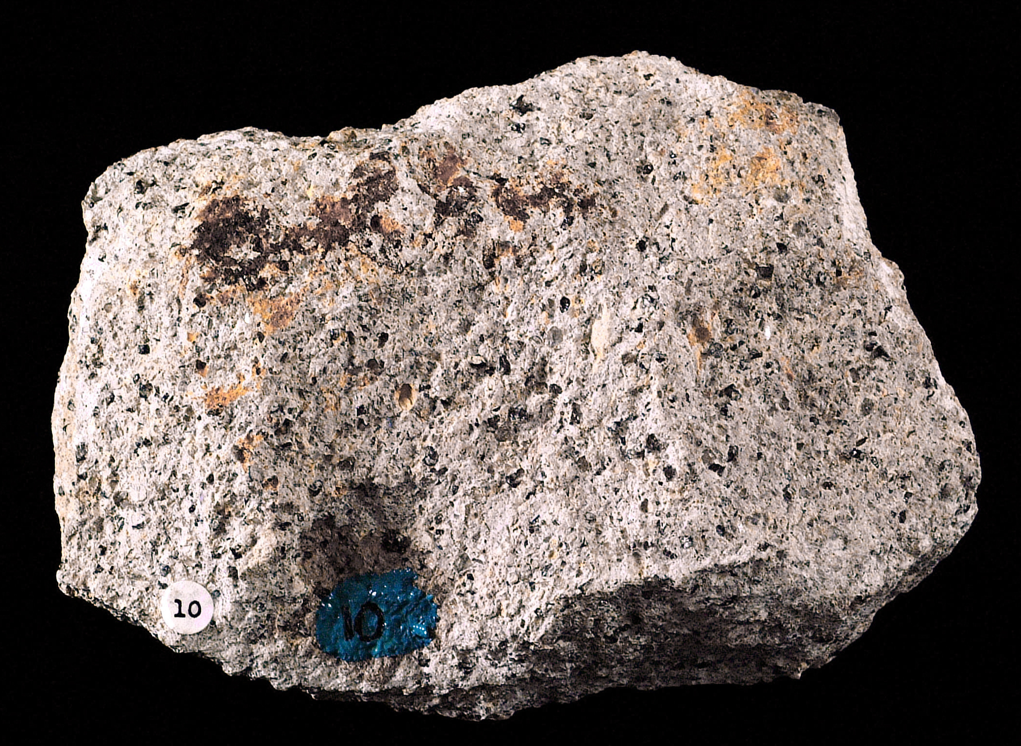
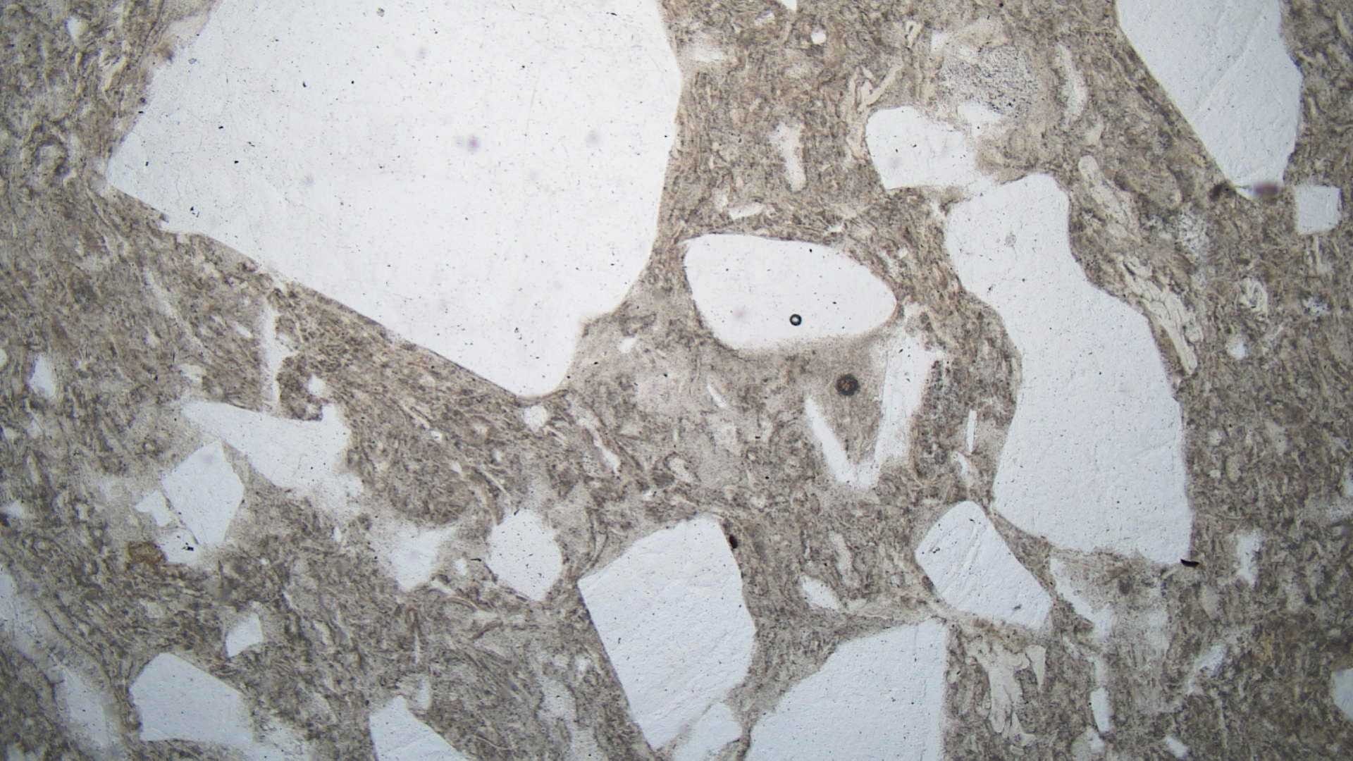
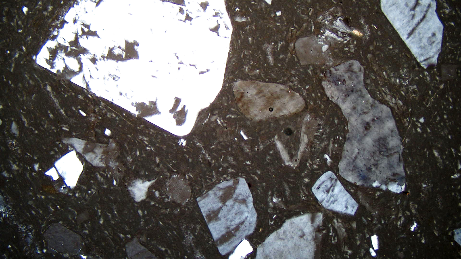
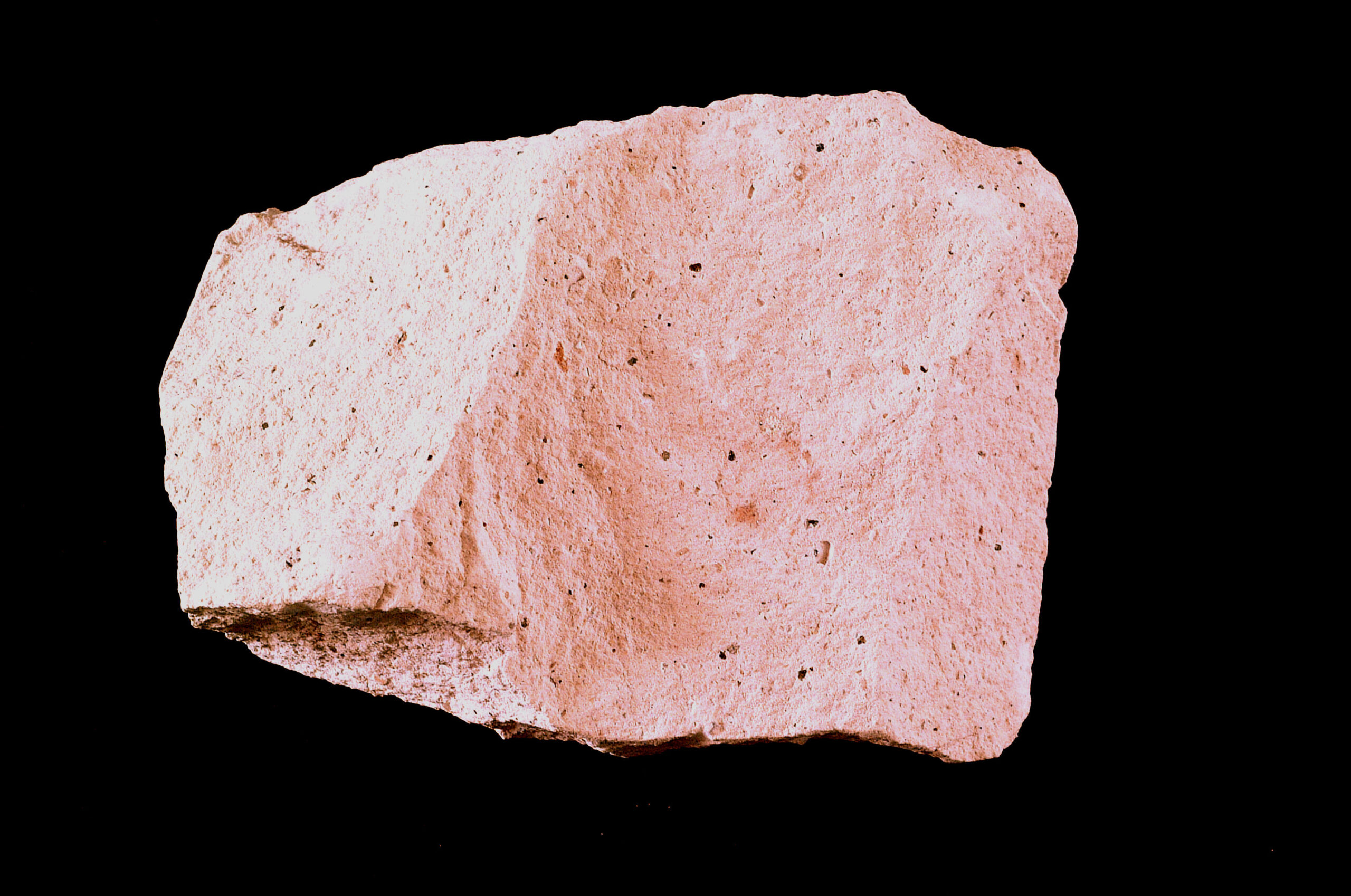
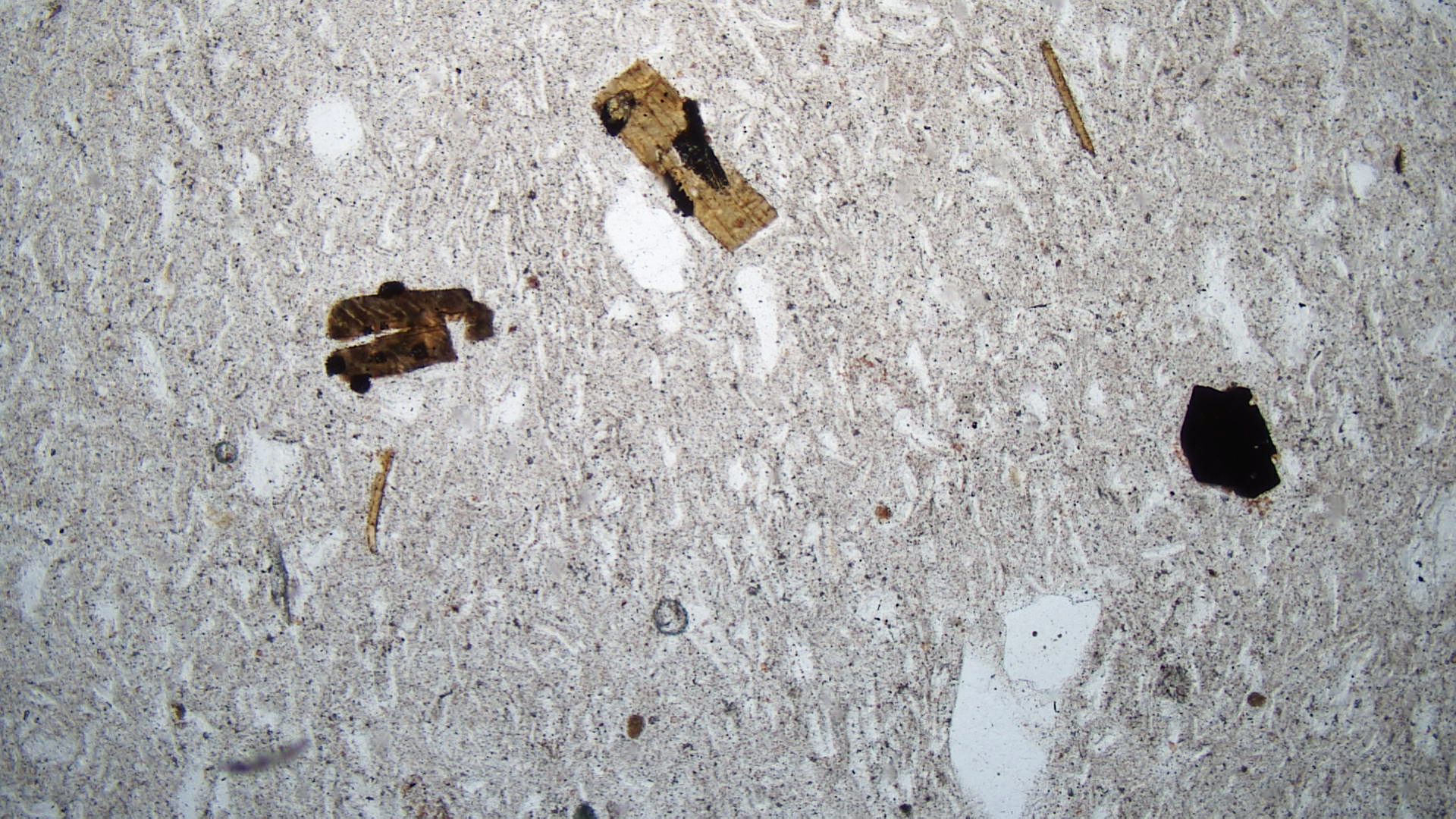
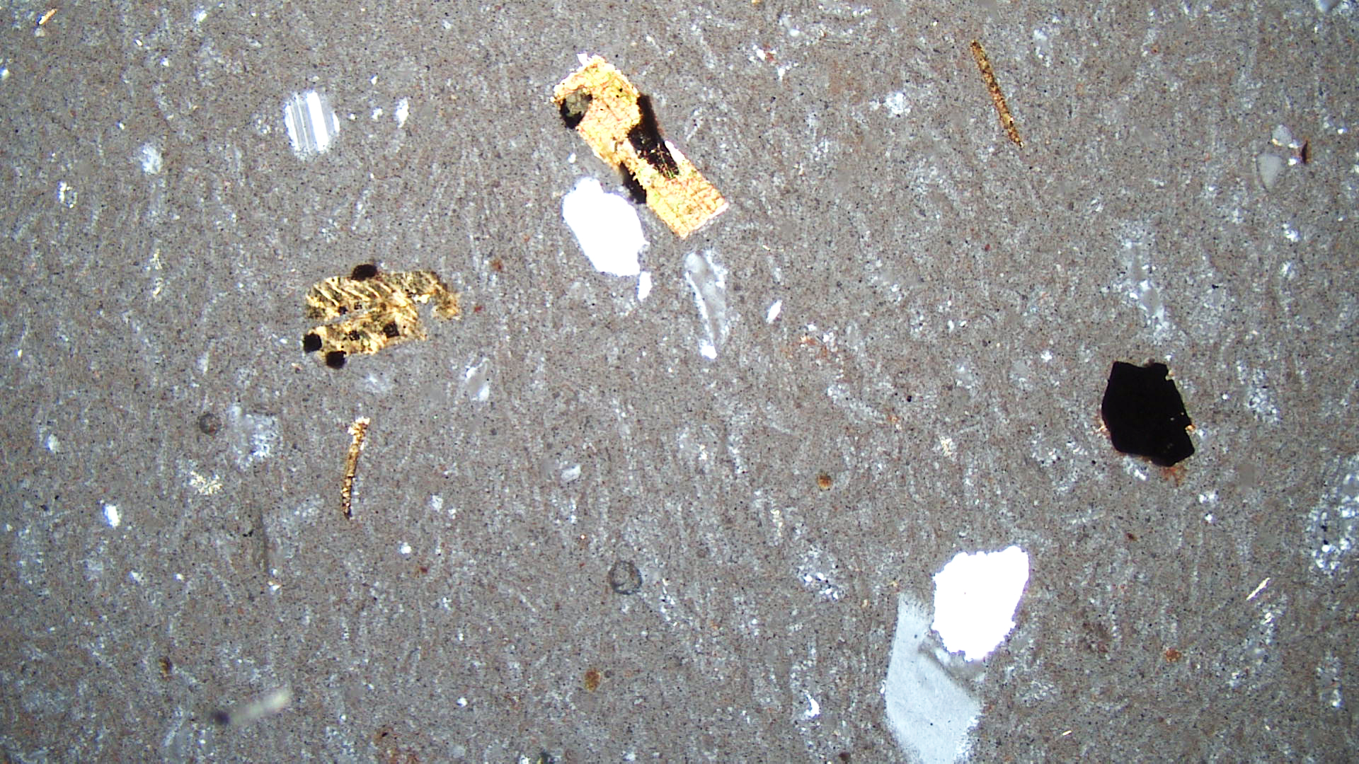
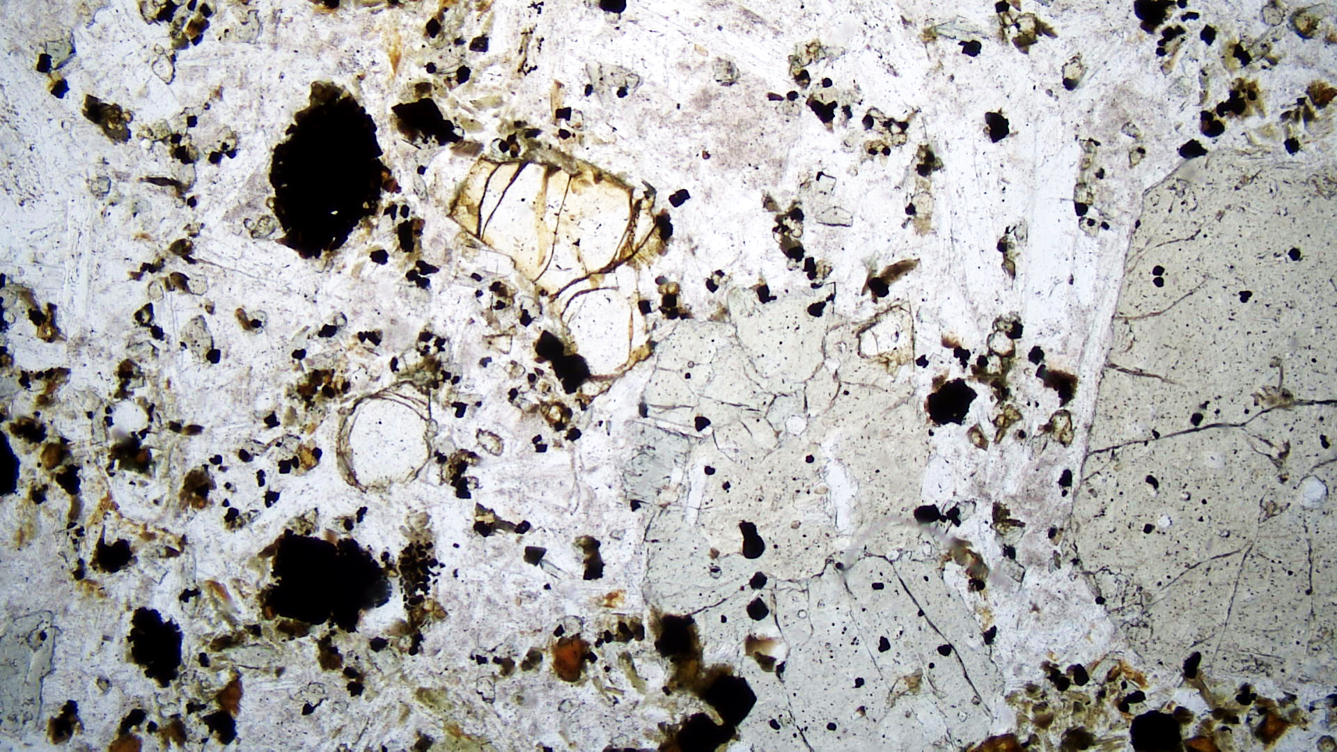
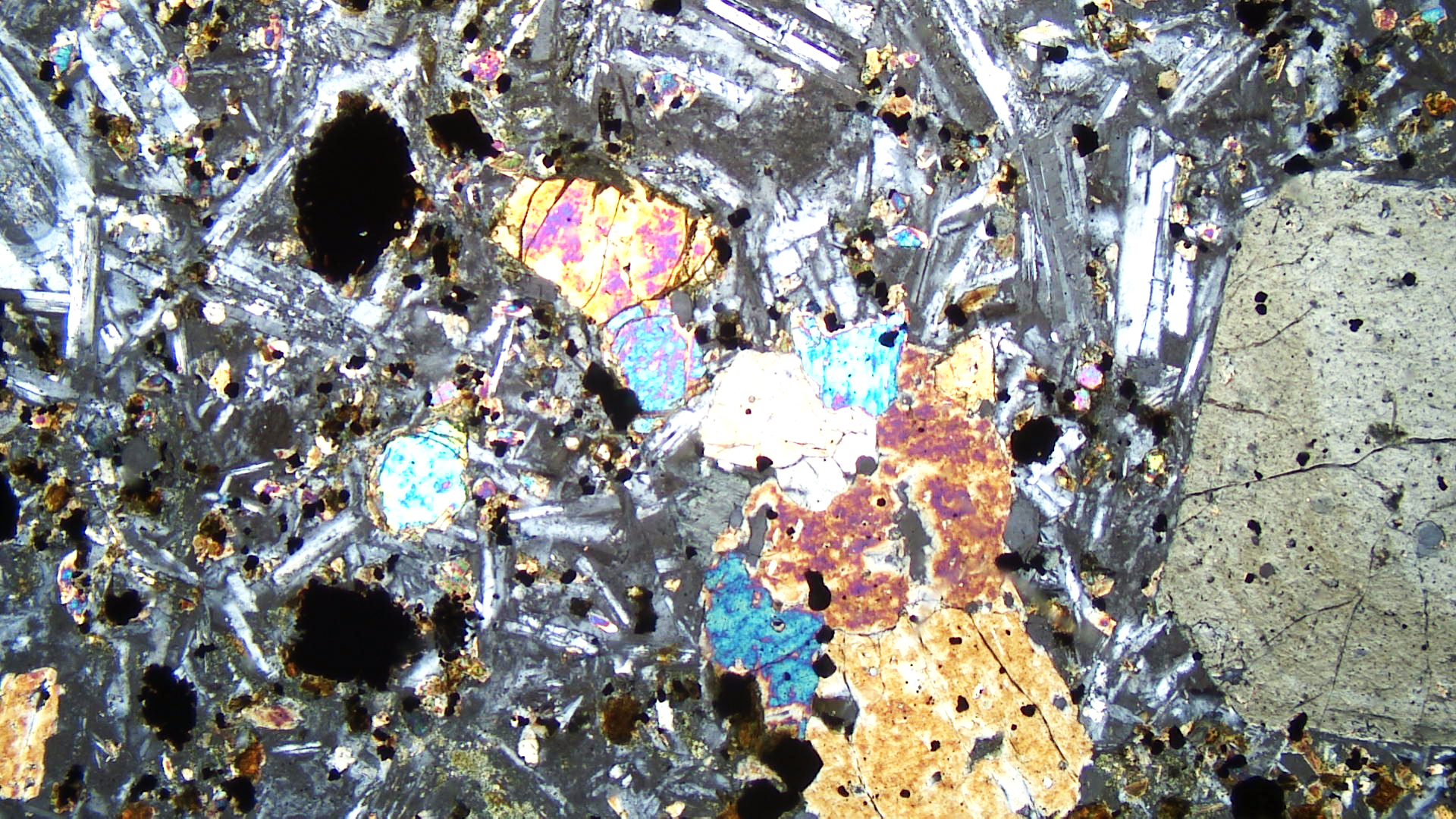
The rhyolite tuff seen in Figure 16.90 is a vesicular tuffaceous fragmental rock from the Frying Pan Basin in Beaverhead County, Montana. It contains 70-80% glass shards and some small scattered phenocrysts of quartz and sanidine in a felsitic groundmass, meaning the groundmass contains extremely small microcrystals of various shapes along with some glass. The small crystals are mostly quartz and sanidine but, in general, are too small to identify. Dark appearing grains in the hand specimen are glassy quartz. Minor hornblende is also present, and vesicles are scattered throughout the sample.
In thin section, feldspar and quartz phenocrysts appear clear (PP).The groundmass is dark colored in the XP view because it contains abundant, slightly devitrified, glass. Although this rock contains small amounts of hornblende, no hornblende is seen in the thin-section photos.
▪Rhyolite Porphyry
|
|
|
|
| ⇒Link to 3D rotatable image of this rhyolite |
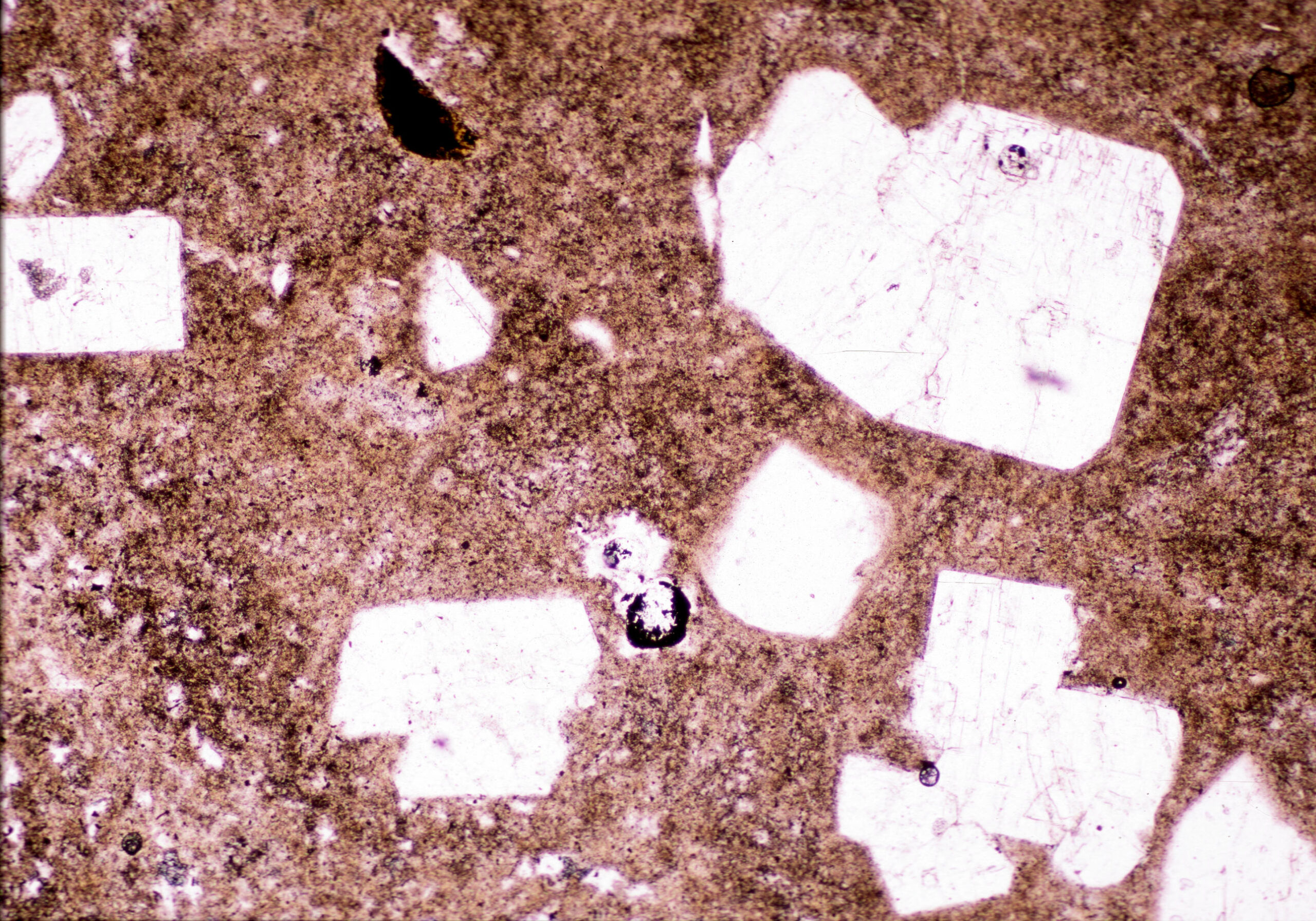
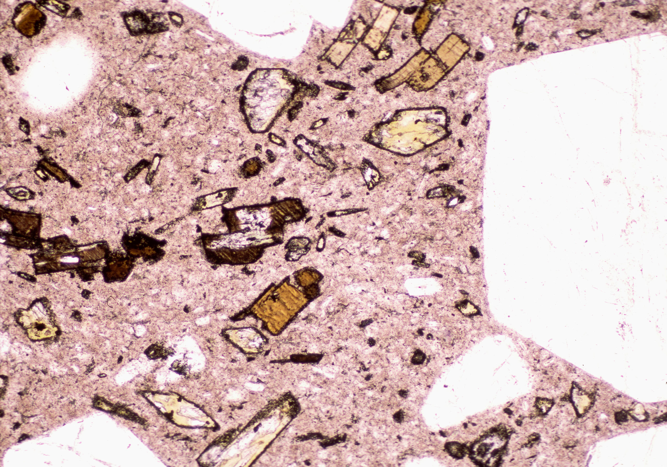
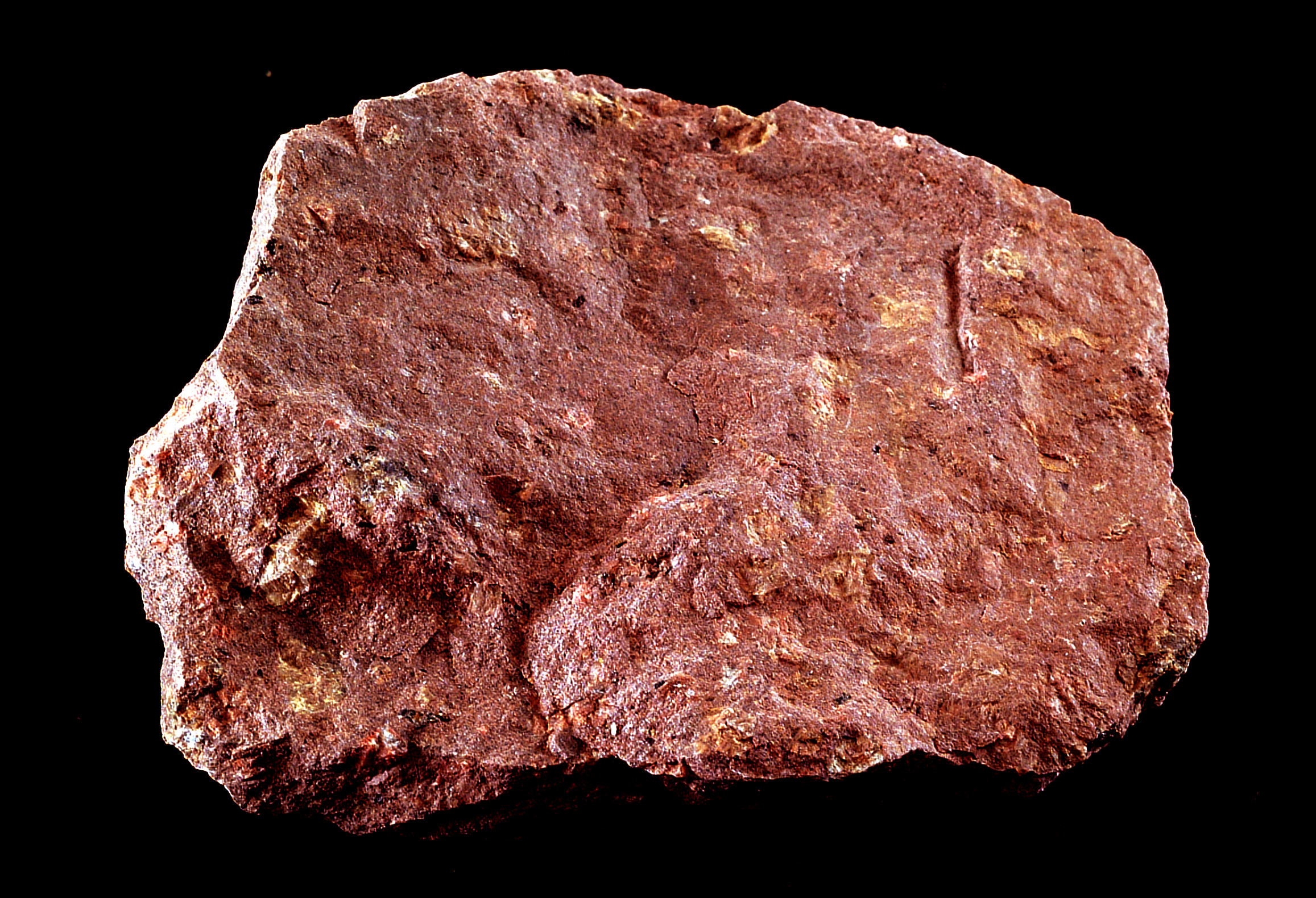
This rhyolite is similar in composition to the previous one, but is more uniform grain-sized, denser, and has less fragmental character. The reddish color is due to staining by hematite. This specimen come from Castle Rock, in Douglas County, Colorado. The slightly vesicular rock has a pyroclastic texture and contains phenocrysts of quartz, sanidine, biotite euhedra, subhedral to euhedral sodic plagioclase laths, a few magnetite crystals, and angular quartz pieces. The small phenocrysts (<1 mm across) appear as dark flecks in the hand sample.
In thin section, the magnetite is opaque, and the biotite appears as brown flakes (PP) and has 2nd-order interference colors. Sanidine, plagioclase, and quartz phenocrysts are clear (PP) and have 1st-order interference colors (XP). The felsitic groundmass is mostly devitrified glass and has white to gray color in the XP view.
▪Rhyolite Porphyry
|
|
|
|


| ⇒Link to 3D rotatable image of this rhyolite porphyry |
This rhyolite porphyry comes from Chafee County, Colorado. The hand specimen is not red-colored like the previous two rhyolite samples because the feldspars are not hematitic. This somewhat vesicular rock contains phenocrysts of smoky quartz up to 5 mm long and slightly smaller subhedral to euhedral phenocrysts of sanidine and oligoclase. The feldspar phenocrysts are hard to see in the hand sample. Scattered dark-colored phenocrysts are mostly quartz.
The thin-section view shows large crystals of blocky sanidine (some with Carlsbad twinning), plagioclase (with albite twins), and (untwinned) quartz in a fine matrix of feldspar, quartz, and glass. Minor magnetite is also present in the groundmass.
▪Dacite
|
|
|
|


| ⇒Link to 3D rotatable model of this dacite |
Dacites contain plagioclase and quartz, commonly with biotite, hornblende, or pyroxenes, and often glass. The dacite seen above comes from northwest of Helena, Montana. It is a porphyritic rock that contains blocky equant to tabular phenocrysts, up to several millimeters across, of whitish plagioclase, dark-gray hornblende and black biotite. The matrix is mostly very fine-grained quartz, feldspar, and glass with a few scatter crystals of biotite and hornblende.
In thin section, felspar and quartz phenocrysts appear clear in the PP view. Hornblende crystals, some with a diamond shape, are light tan. Biotite appears as brown parallel-sided flakes. The matrix includes opaque magnetite, minor brown biotite, and greenish hornblende (PP). The XP view shows zoned plagioclase grains, some displaying twinning. Biotite’s brown color masks its interference colors. The altered hornblende has up to 2nd-order colors. The matrix is nearly isotropic because of abundant glass.
16.2.2 Relatively SiO2-poor Volcanic Rocks
▪Trachyte Porphyry
|
|
|
|

| ⇒Link to 3D rotatable image of this trachyte porphyry |
This porphyritic trachyte, stained red by hematite, comes from Essex County, New York. Trachytes are composed mostly of alkali feldspar with minor amounts of mafic minerals. They are volcanic equivalents of the plutonic rock syenite. Most trachytes are porphyritic, containing phenocrysts of sanidine or orthoclase and, less commonly, other minerals. Orthoclase phenocrysts in this rock are quite small but some can be seen if you enlarge the hand-sample photo – they are slightly less red than other parts of the rock. Trachytes commonly have a felsitic texture, which means the groundmass contains extremely small microcrystals of various shapes along with some glass.
The thin-section views show a large of turbid orthoclase phenocryst near the left edge, with many small feldspar crystals in the matrix. Clear areas in plane-polarized light are glass; the glass is isotropic in the crossed-polars view. The matrix also contains some small altered and unidentifiable mafic grains.
▪Trachyte Porphyry
|
|
|
|
| ⇒Link to 3D rotatable image of this trachyte porphyry |
This trachyte porphyry comes from near Cripple Creek, Teller County, Colorado. It contains millimeter-sized phenocrysts of sanidine and smaller phenocrysts of hornblende. Both are highly altered. The matrix contains mostly plagioclase, altered sanidine and lesser amounts of quartz.
In thin section, the feldspar is clear (PP) and zoned (XP). It is significantly replaced by kaolinite. Small dark-brown grains with high relief are altered mafic minerals. The equigranular and glassy matrix (probably) contains plagioclase and K-feldspar but is nearly isotropic.
▪Latite Porphyry
|
|
|
|


| ⇒Link to 3D rotatable model of this latite porphyry |
Latites contain subequal amounts of plagioclase and K-feldspar with minor or no quartz. This sample is a latite porphyry from Montana’s Bearpaw Mountains. The hand sample contains conspicuous tabular phenocrysts of white plagioclase up to several millimeters long. Smaller flakes of biotite are also present but hard to see. Minor pale green augite is present but cannot be seen in the photo.
The thin section (PP) shows a mostly clear groundmass composed of a mix of feldspars, small grains of biotite, and specks of opaque magnetite. Larger laths of clear plagioclase, and greenish biotite and augite are also present. In crossed polars, twinned plagioclase has typical 1st-order colors. The altered mafic minerals have 2nd-order green interference colors. Minor magnetite can also be seen.
▪Hornblende Andesite
|
|
|
|

| ⇒Link to 3D rotatable model of this hornblende andesite |
Andesites contain plagioclase, lesser amounts of quartz and K-feldspar, and accessory mafic minerals. This andesite, from Mt. Shasta in California contains black hornblende and augite crystals in a lighter-gray groundmass composed of fine plagioclase, augite, magnetite, and glass. The augite crystals appear as dark laths up to several millimeters long in the hand-sample photo.
In thin section, twinned plagioclase is mostly clear and has 1st-order interference colors. Augite is also mostly clear but, in contrast with the plagioclase, has upper 2nd-order interference colors. Small dark-colored hornblende crystals have altered to pyroxene and magnetite. The glassy groundmass is opaque.
▪Amygdaloidal Basalt
|
|
|
|
Basalt is the most common kind of volcanic rock. All basalts are fine-grained, containing essential plagioclase and pyroxene, typically augite. Quartz, hornblende, biotite, orthopyroxene, and feldspathoids may be present as accessory minerals.
The hand sample of basalt seen above is a very altered amygdaloidal olivine basalt from Michigan’s Keweenaw Peninsula. The millimeter-sized amygdules, which are slightly lighter colored than the fine material around them, contain quartz, epidote, calcite, and chlorite. The primary mafic minerals in this rock have been replaced by secondary chlorite, serpentine, magnetite, and hematite.
The large amygdule in the upper right of the thin-section views contains quartz with other unidentifiable minerals. The groundmass contains small laths of clear plagioclase with low-order interference colors. Mafic minerals are highly altered. Light-green grains were once clinopyroxene, now replaced by chlorite. Darker altered grains, stained red, were once olivine. Abundant secondary magnetite is present.
▪Basalt
Clinopyroxene and plagioclase are essential minerals in basalts. The fine-grained basalt seen below, from Chimney Rock Park in Somerset County, New Jersey, contains only these minerals, along with volcanic glass that is partly devitrified. The hand sample has a uniform dark color and is so fine grained that no individual crystals can be seen.
|
|
|
|
In the thin-section PP view, one large phenocryst dominates the right side of the photo. Finer groundmass material dominates the left side. The groundmass contains small laths of clear plagioclase, small equant clinopyroxene (augite) grains that have a faint hint of green color, and dark brown/black glass that appears nearly opaque. Some glass grains have altered to become light-brown/tan chlorite.
In crossed polars, the large phenocryst is revealed to be an agglomeration of multiple mineral grains – mostly augite. This is a classic example of a glomeroporphyritic texture. Plagioclase has typical twinning and 1st-order white to black interference colors; augite displays up to 3rd-order colors. Most of the glass is isotropic black.
▪Olivine Basalt Porphyry
Clinopyroxene and plagioclase are essential minerals in basalts, but other mafic minerals are commonly present as accessories. The medium-grained basalt seen below, from Boulder County, Colorado, contains phenocrysts of augite, olivine, and magnetite surrounded by plagioclase. In the hand-sample view, several prominent black euhedral to subhedral augite phenocrysts are larger than other minerals in the rock. Olivine crystals are hard to pick out from surrounding plagioclase but have a slightly greenish hue.
|
|
|
|
| ⇒Link to 3D rotatable model of this basalt porphyry |
In thin section under plane polarized light, the clear grains are mostly laths of plagioclase. Augite appears light green, and olivine is generally clear with high relief. Magnetite grains are opaque. In the XP view, plagioclase has 1st-order interference colors, augite has up to high 2nd-order interference colors, and olivine has patchy interference colors up to 3rd order. The large augite grain on the right with 1st-order interference colors displays concentric zoning.
16.2.3 Feldspathoid-Bearing Volcanic Rocks
▪Phonolite
|
|
|
|
| ⇒Link to 3D rotatable image of this phonolite |
Phonolites are rare volcanic rocks rich in nepheline and K-feldspar. This phonolite is from Cripple Creek, Colorado. Although difficult to see in hand specimen, it contains feldspars (anorthoclase phenocrysts) and nepheline as major minerals, in a finer-grained glassy matrix that contains orthoclase.
In thin section, the large clear grains are nepheline. Finer clear grains in the matrix are feldspar in a sea of glass (PP). In the XP view, feldspar and nepheline have 1st-order interference colors, and the matrix is mostly isotropic. Minor clinopyroxene (aegirine) is present but difficult to discern.
▪Tephrite
|
|
|
|

| ⇒Link to 3D rotatable image of this tephrite |
Tephrites are extrusive rocks that contain calcic plagioclase, clinopyroxene and lesser amounts of feldspathoids. The hand sample seen above is a highly vesicular tephrite from a 2-km wide caldera called the Laacher See, in Germany. The rock contains black phenocrysts of augite in a glassy matrix. Plagioclase and leucite are also present but too fine-grained to pick out.
The thin-section photos are of a different tephrite, this one from Mt. Vesuvius, Italy. The PP view shows large rounded white leucite crystals, greenish-gray augite phenocrysts, and small clear plagioclase laths in a glassy groundmass. In the XP view, plagioclase has 1st-order interference colors and pyroxene has up to high 2nd-order interference colors. Leucite has very low-order interference colors, appearing almost isotropic. The matrix is mostly black (isotropic) glass except for small laths of plagioclase.
▪Leucitite
|
|
|
|


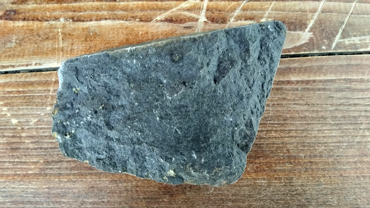
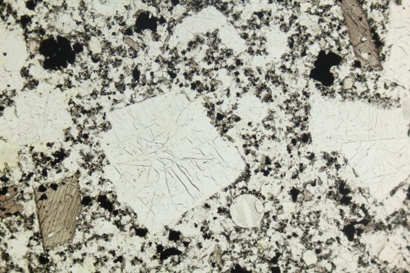
Leucitites contain mostly leucite and clinopyroxene. The hand sample above is a leucitite from Mt. Vesuvius, Italy. The thin-section views are of a different leucitite from Vulsini, Italy, about 300 km northwest of Mt. Vesuvius.
The thin section contains several greenish clinopyroxene phenocrysts – one large and elongate – with smaller leucite phenocrysts (PP). In the XP view, because of its very low birefringence, the leucite crystal appear nearly isotropic. The large zoned and twinned pyroxene displays high 2nd-order blue-green interference colors, and the smaller pyroxene grains have lower 2nd-order interference colors. Finer grains in the groundmass are a mix of leucite and pyroxene.
▪Nephelenite
|
|
|
|
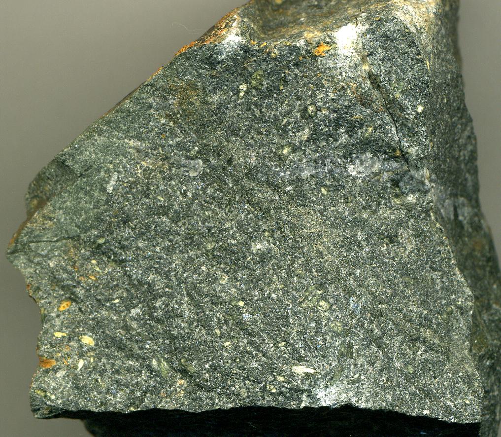
Nephelinites contain mainly augite, nepheline, and glass. The hand sample seen above is a nephelinite from the Czech Republic. It is fine-grained with a uniform texture. The thin-section views are of a different nephelinite from Cape Verde.
In the thin section PP view, large blocky nepheline phenocrysts are nearly clear and smaller augite crystals are greenish-gray. Some large opaque magnetite grains are also present. In the XP view, nepheline has 1st-order interference colors and the pyroxene up to high 2nd-order interference colors. The glassy matrix is nearly isotropic but contains small grains of nepheline and augite.
16.3 Metamorphic Rocks
16.3.1 Granitic gneisses
▪Augen Gneiss
|
|
|
|

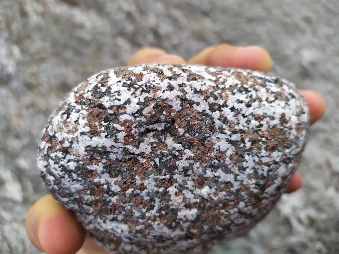
The granitic hand specimen above is an augen gneiss from the western Adirondack Mountains, New York. It contains augen (“eyes”) of pink K-feldspar, plagioclase, and quartz with biotite at their centers.
The clear minerals in the PP thin-section view are quartz and feldspars. The darker colored grains are shredded biotite mixed with hornblende. In the XP view, quartz and the two feldspars can be distinguished based on absence or presence of twinning. The biotite appears to be part way to becoming all hornblende. Although the morphology of some grains looks like biotite, the interference colors are all low-order, suggesting alteration. Some of the former biotite displays typical hornblende green interference colors.
▪Granitoid Gneiss
|
|
|
|

This granitic gneiss, from Salisbury, North Carolina, looks like a granite but displays a distinct foliation – pink K-feldspar rich layers alternate with whiter quartz-rich layers. Scattered magnetite and other dark-colored mineral grains are present but hard to see.
Most of the thin-section view is feldspar (plagioclase with some microcline) and quartz. These minerals look the same in plane polars but can be distinguished in crossed polars because the quartz is untwinned, the plagioclase displays albite twins (somewhat distorted), and the K-feldspar display microcline twins. All three display undulatory extinction due to strain on the rock. Muscovite and epidote, along grain boundaries, has upper 2nd-order and higher-order interference colors.
▪Biotite Gneiss
|
|
|
|


This biotite gneiss comes from Uxbridge, Massachusetts. The leucocratic layers are entirely quartz and K-feldspar that alternate with stringers containing mostly black biotite.
The thin-section PP view shows thin stringers of brown biotite separated by clear quartz and K-feldspar. The feldspar grains have a rougher surface than the quartz grains due to incipient alteration. In crossed polars, quartz has undulatory extinction, and the K-feldspar displays hazy microcline twinning. Both have low 1st-order interference colors. Biotite has 2nd-order colors, partially masked by the color of the mineral, and has a typical pebbly surface texture. A few very small zircon grains display high-order interference colors. Although not seen here, other parts of the thin section contain minor hornblende.
16.3.2 Metamorphosed Mafic and Ultramafic Rocks
Photos in this section show typical metamorphosed mafic/ultramafic rocks. Protoliths for these rocks were, for the most part, basalts, although some protoliths may have been ultramafic rocks, or may have been plutonic rocks. The photos begin with serpentinite – a very low-grade metamorphic rock . Subsequent photos show rocks of increasing grade, ending with a garnet granulite.
▪Serpentinite
|
|
|
|
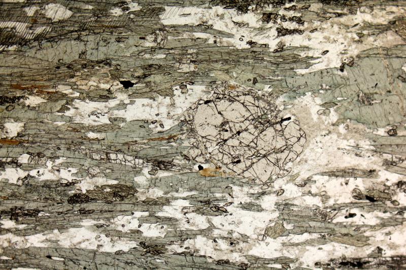
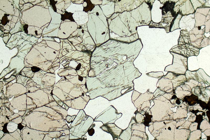
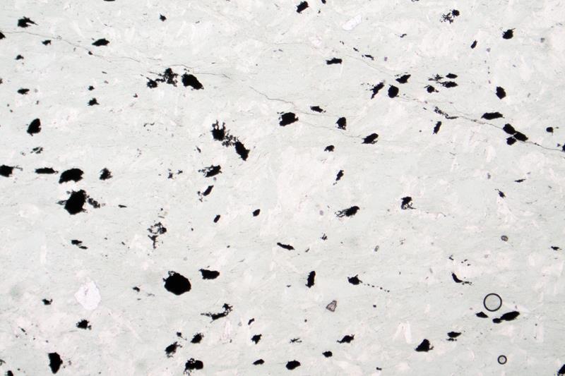
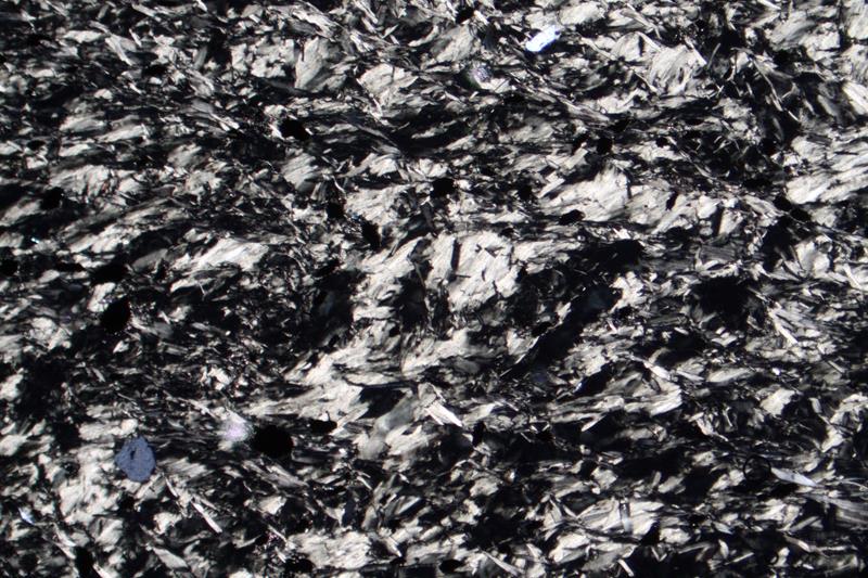
Serpentinite is a general name for metamorphic rocks dominated by minerals of the serpentine group, most often antigorite or chrysotile. These rocks are products of serpentinization, which is very low-grade, mostly hydrothermal, metamorphism of ultramafic protoliths.
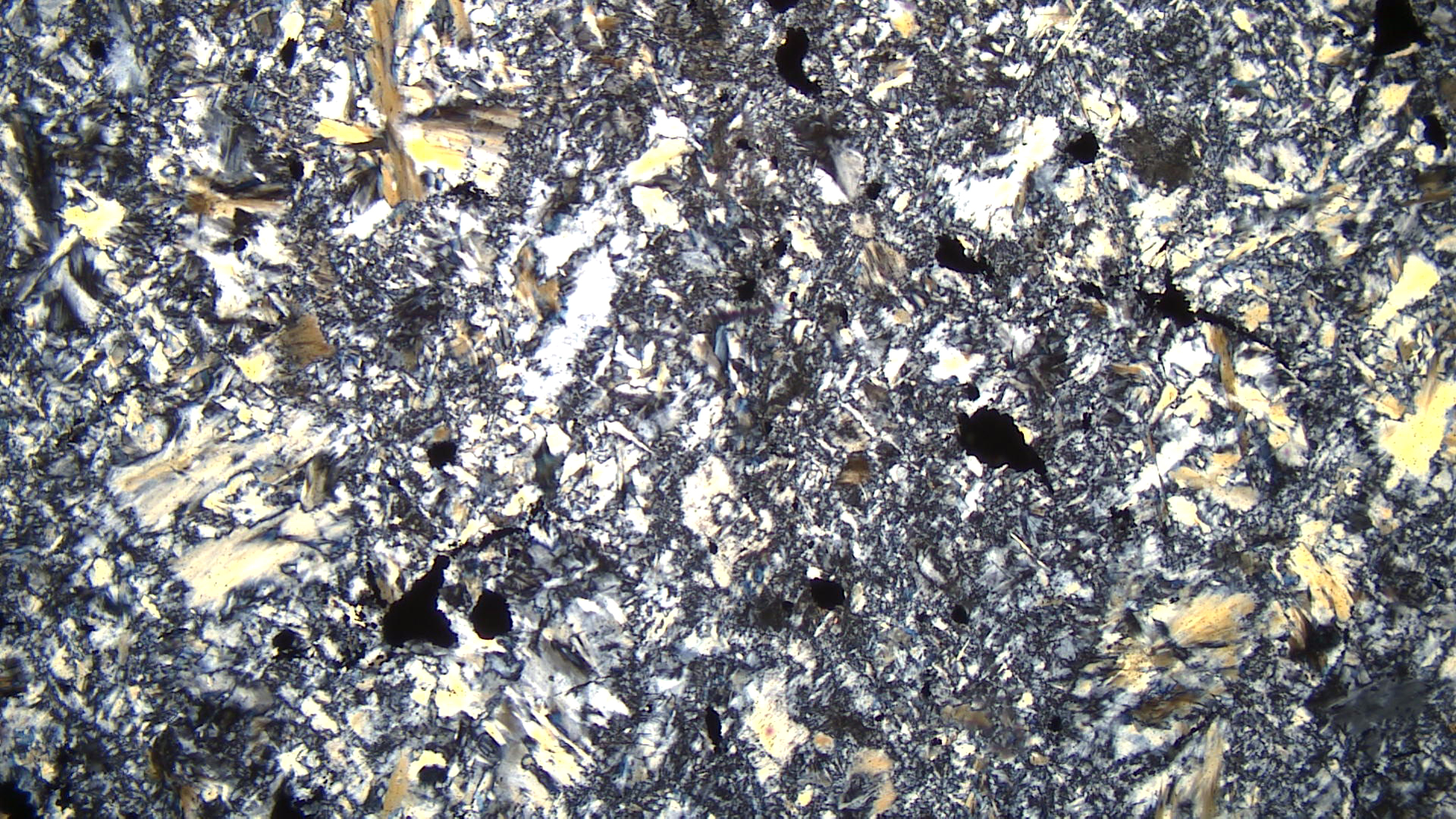
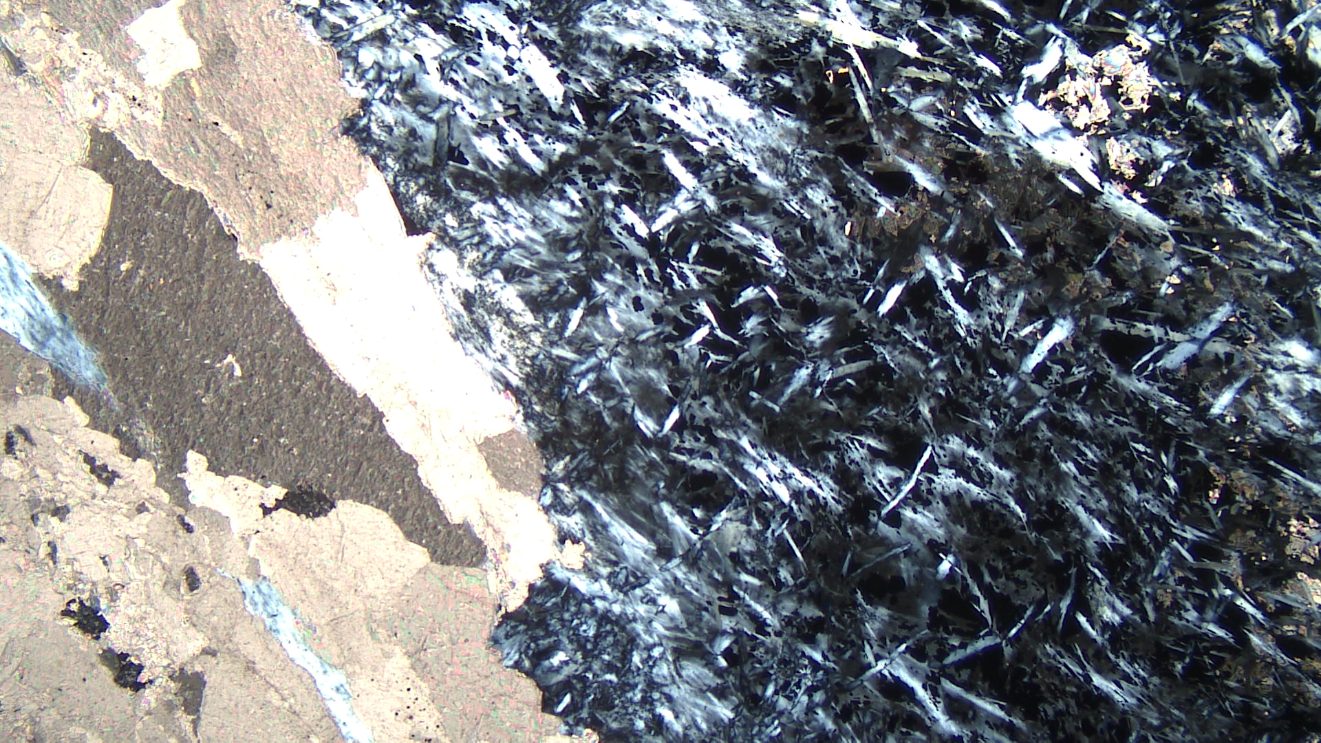
The three photos above are of a serpentinite from near Eden Mills in northern Vermont. The hand sample looks quite fresh, but the thin-section views reveal that the rock, which originally contained olivine and pyroxene, is now almost entirely serpentine. Opaque magnetite grains are common in serpentinites and can be seen in the thin-section. In crossed polars, two different kinds of serpentine are apparent. Some, which displays up to 1st-order yellow interference colors, is arrayed in radiating fibrous splays. Most of the serpentine, however, is in a fine-grained groundmass that has only lower 1st-order white to gray colors.
▪Verde Antique (Serpentinite)
|
|
|
|
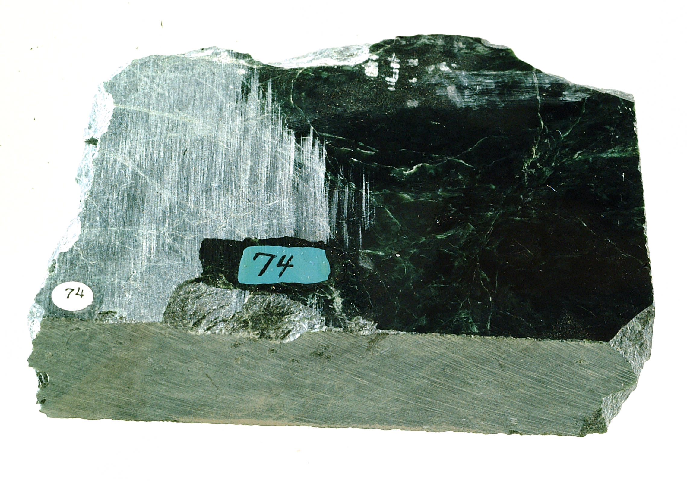
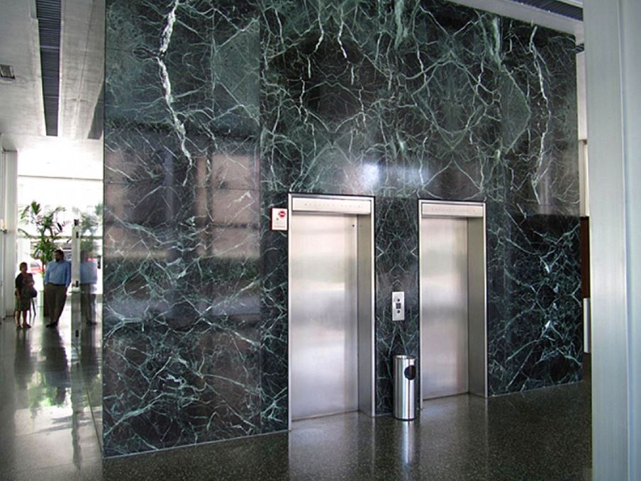
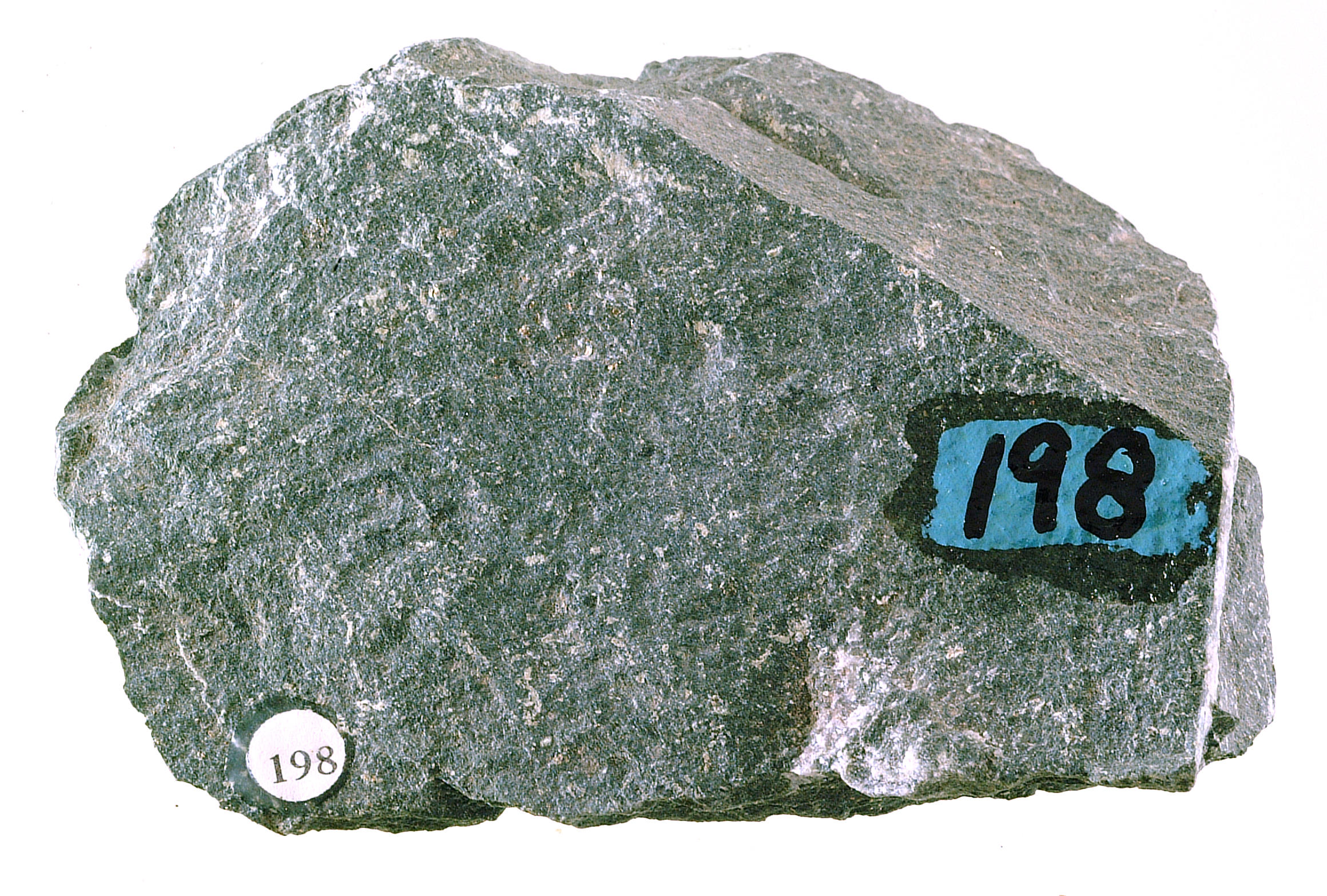
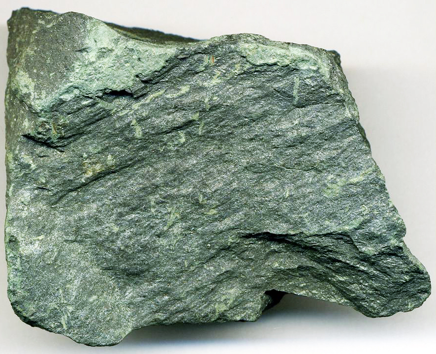
Verde antique is a type of serpentinite breccia that can be polished and used as a building stone. It is sometimes thought of as a variety of marble because it is used for the same purposes as marble, but it is not a kind of marble – it is a metamorphosed ultramafic rock. The sample seen in the three photos above comes from near Rochester in central Vermont. The photo in Figure 16.150 shows slabs of polished verde antique decorating an office building lobby.
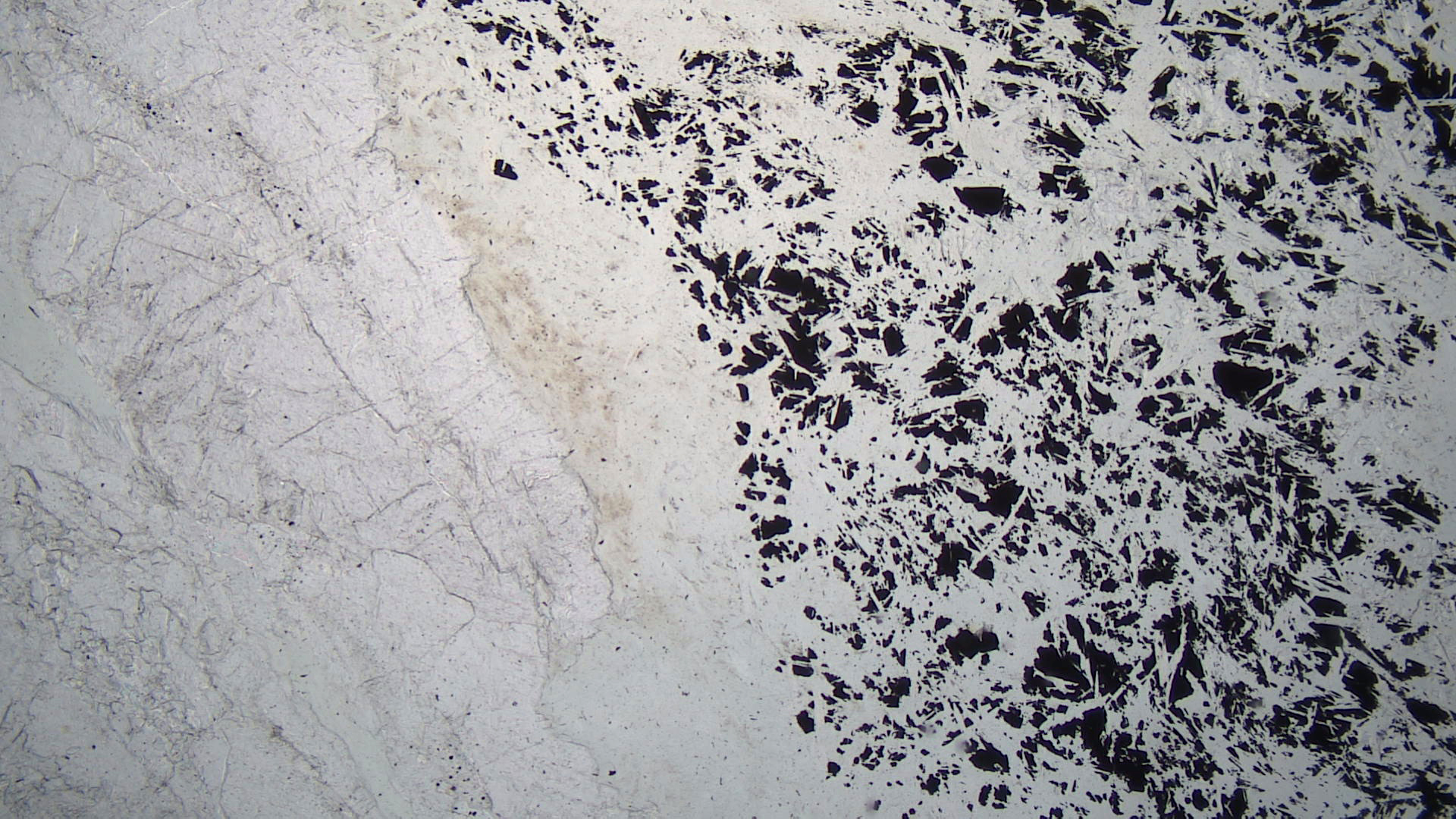
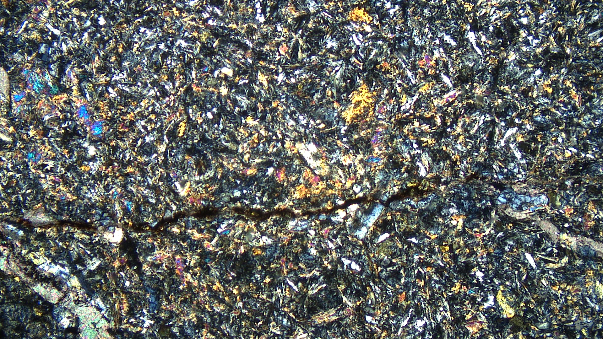
The plane-polarized thin-section view shows very light-green serpentine speckled with opaque magnetite on its right side. The magnetite is why the rock has such a dark color. The left side of the thin section is entirely carbonate (magnesite). In crossed polars, the serpentine has its standard 1st-order interference colors and the carbonate has very high-order pastel colors.
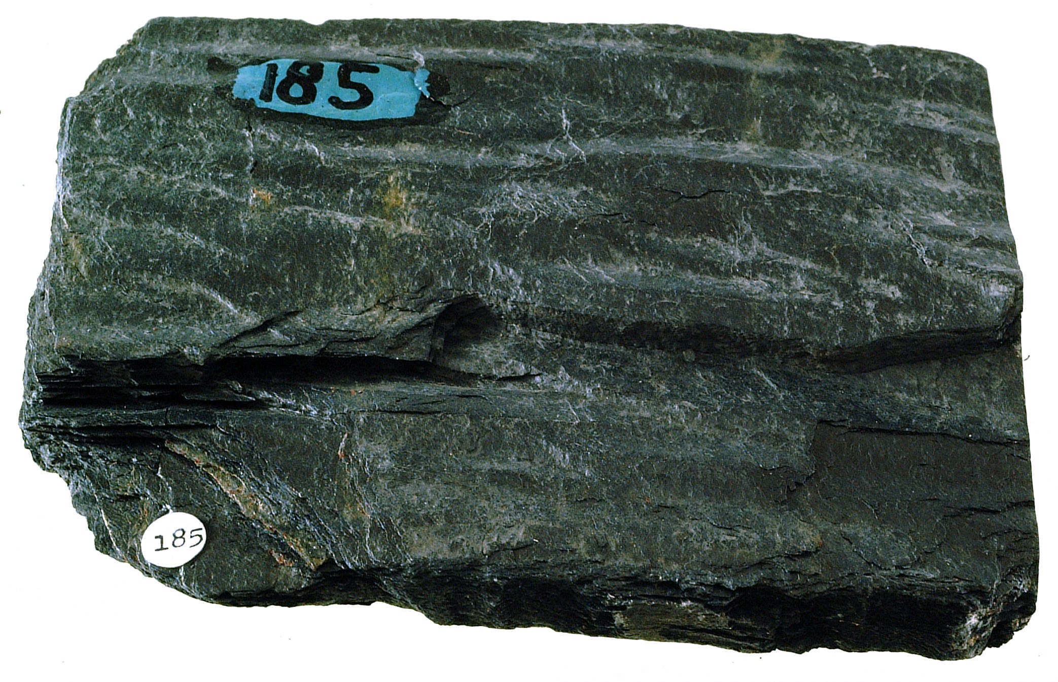
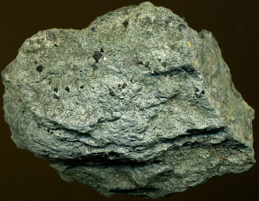
▪Greenstone
|
|
|
|

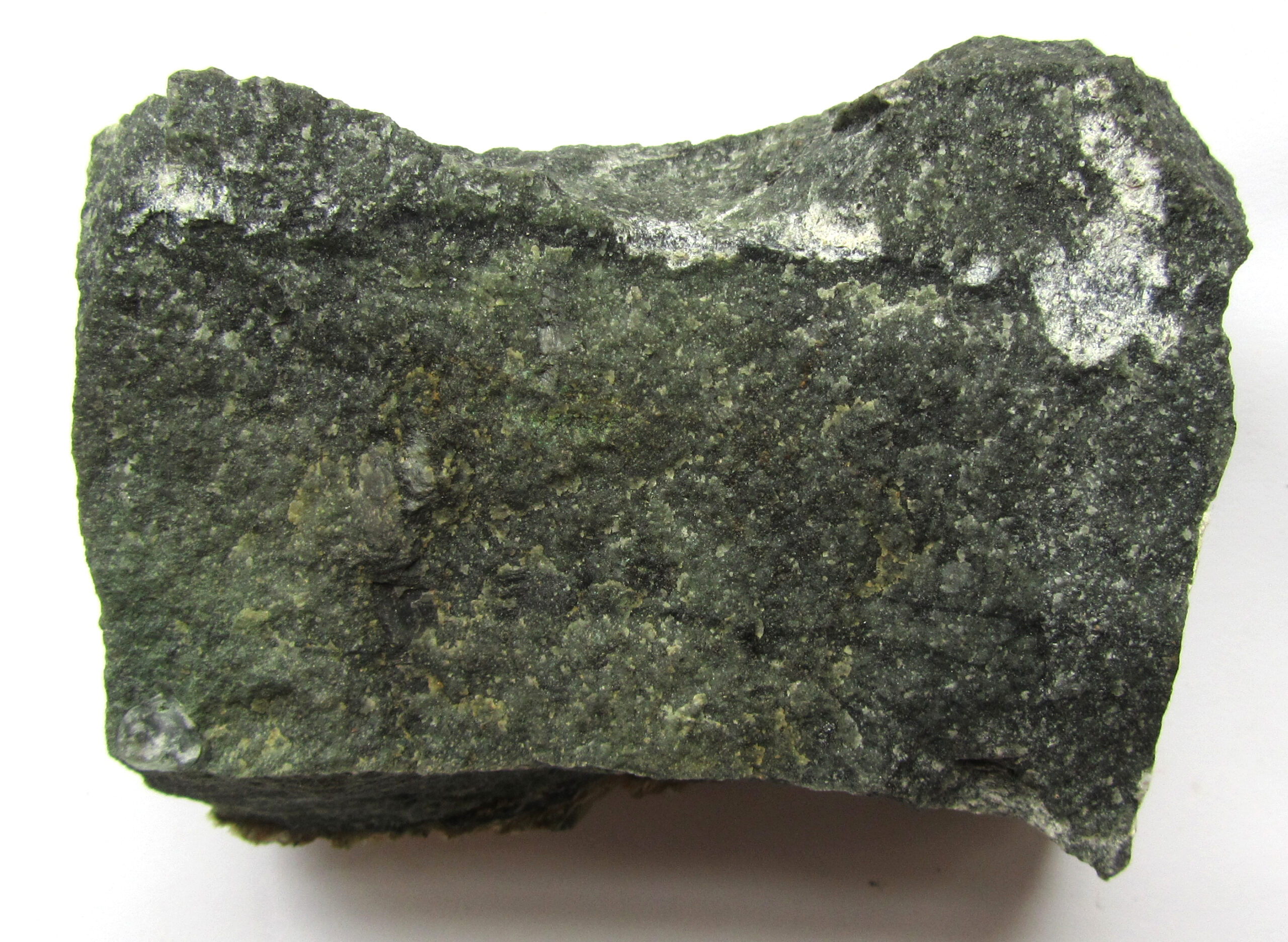
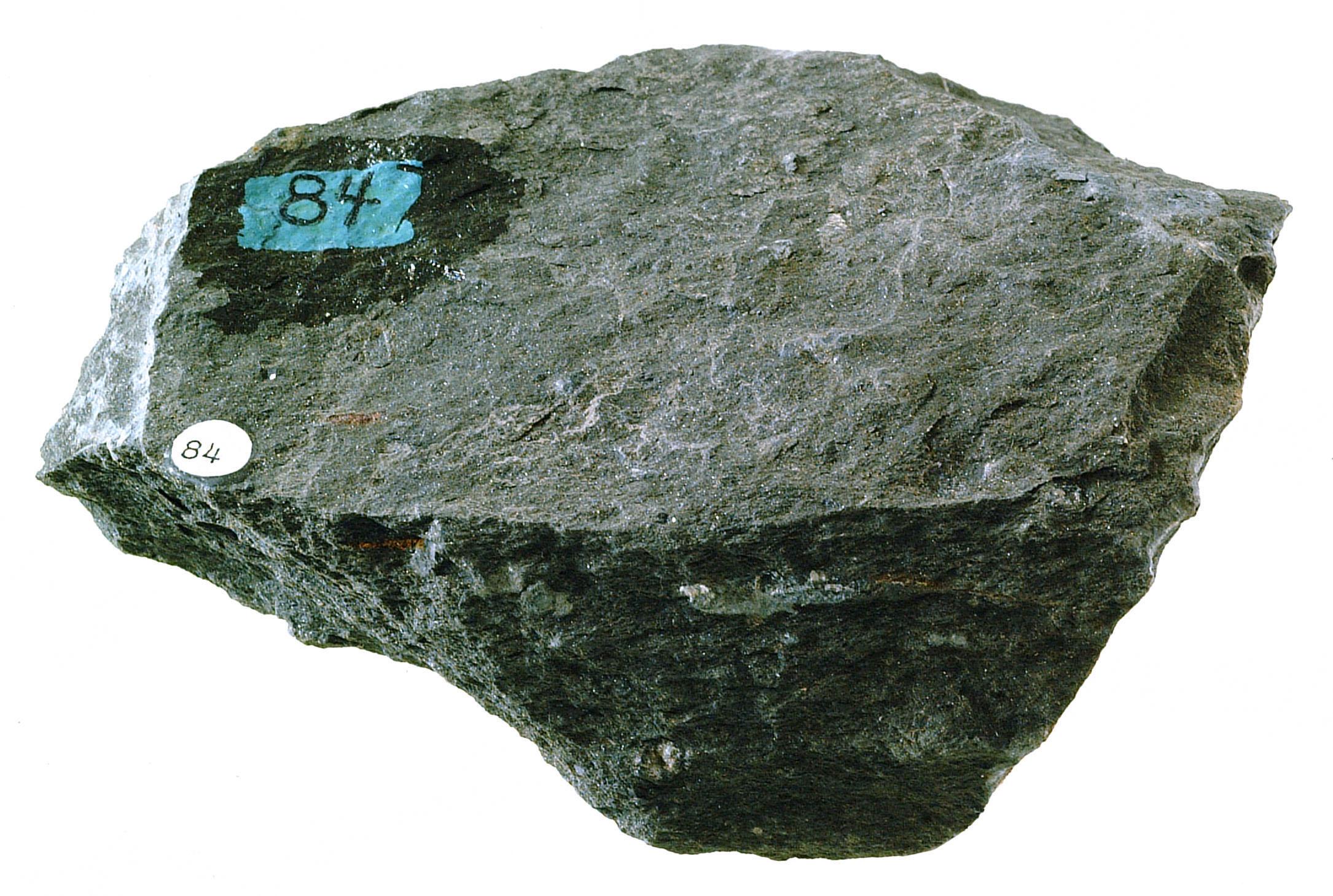
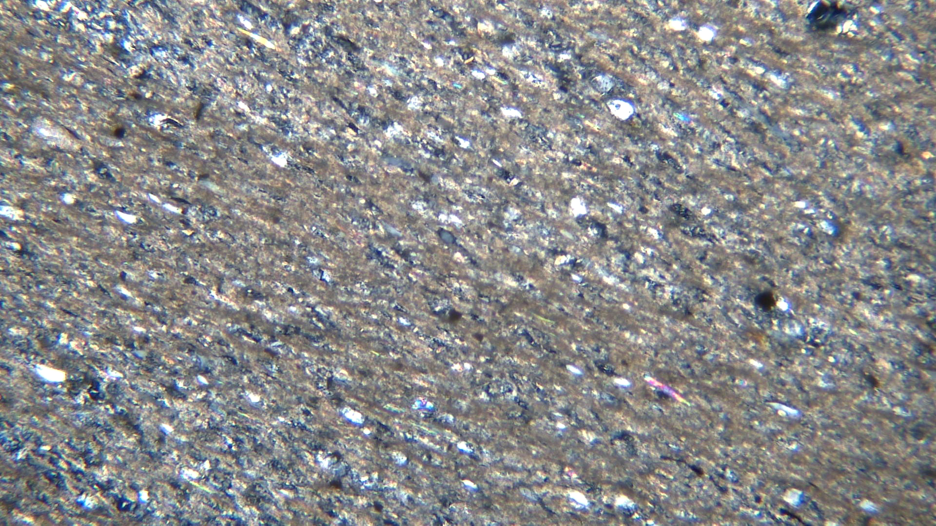
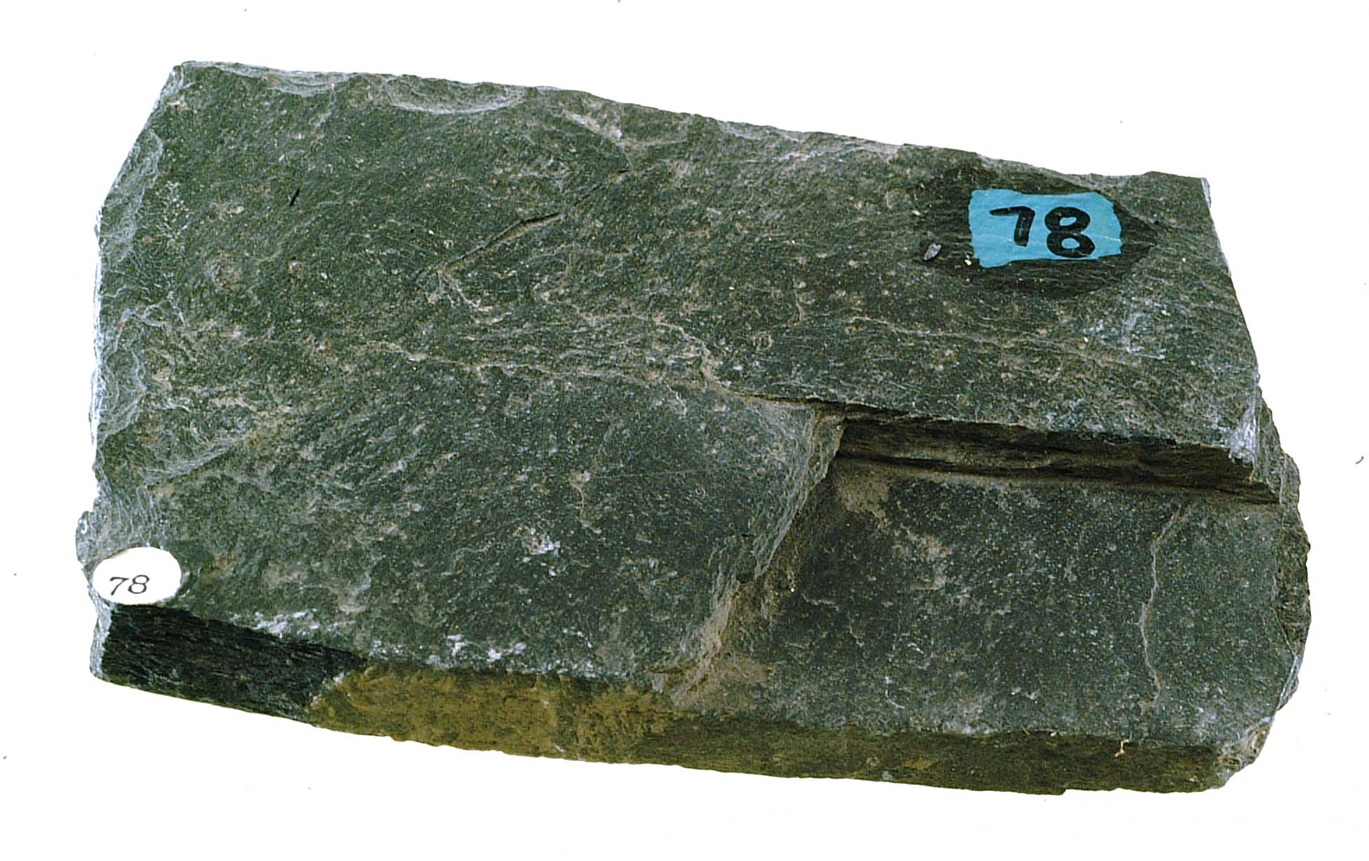
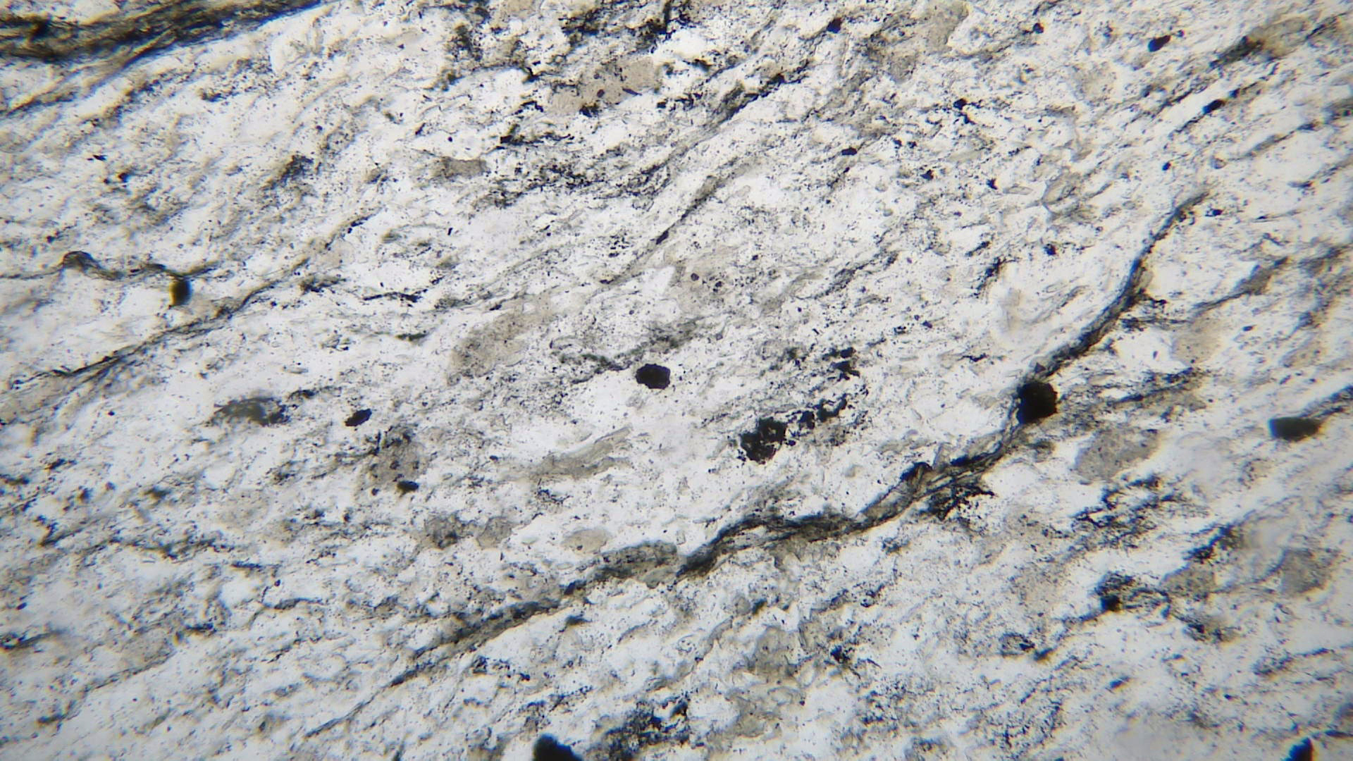
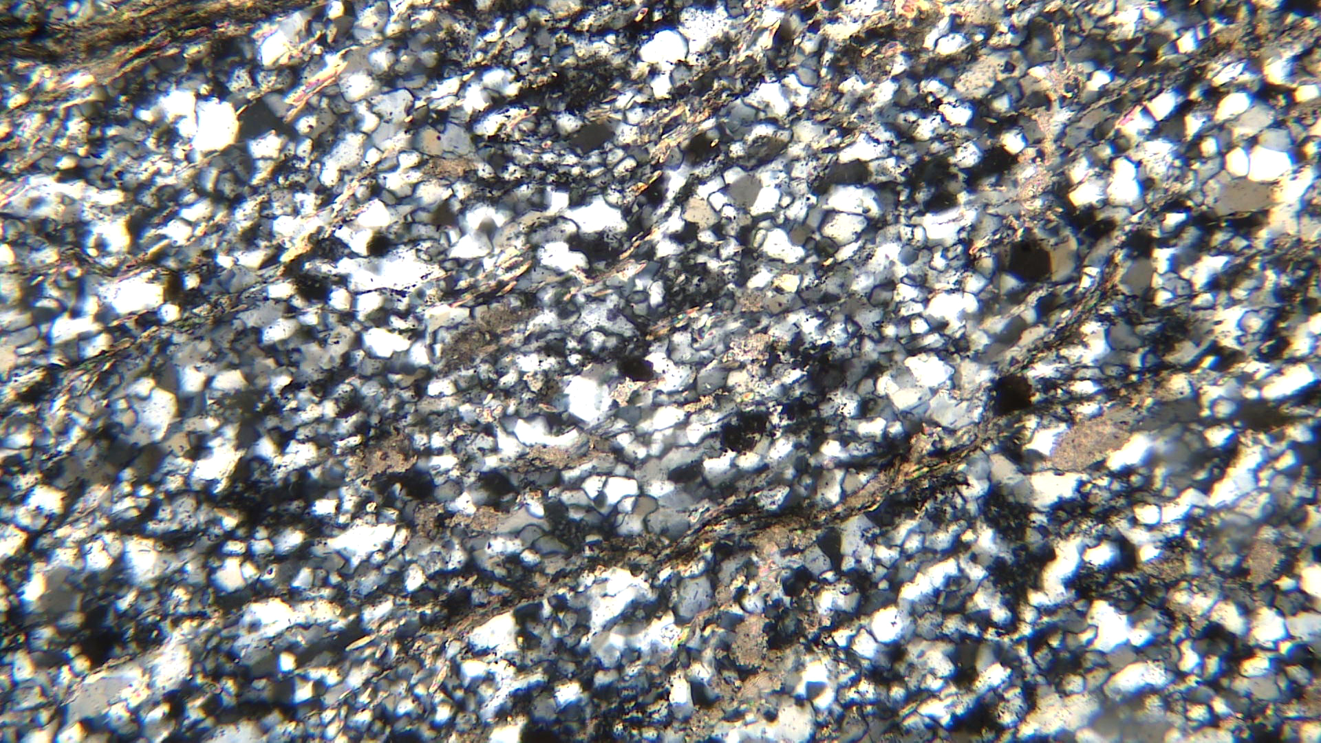
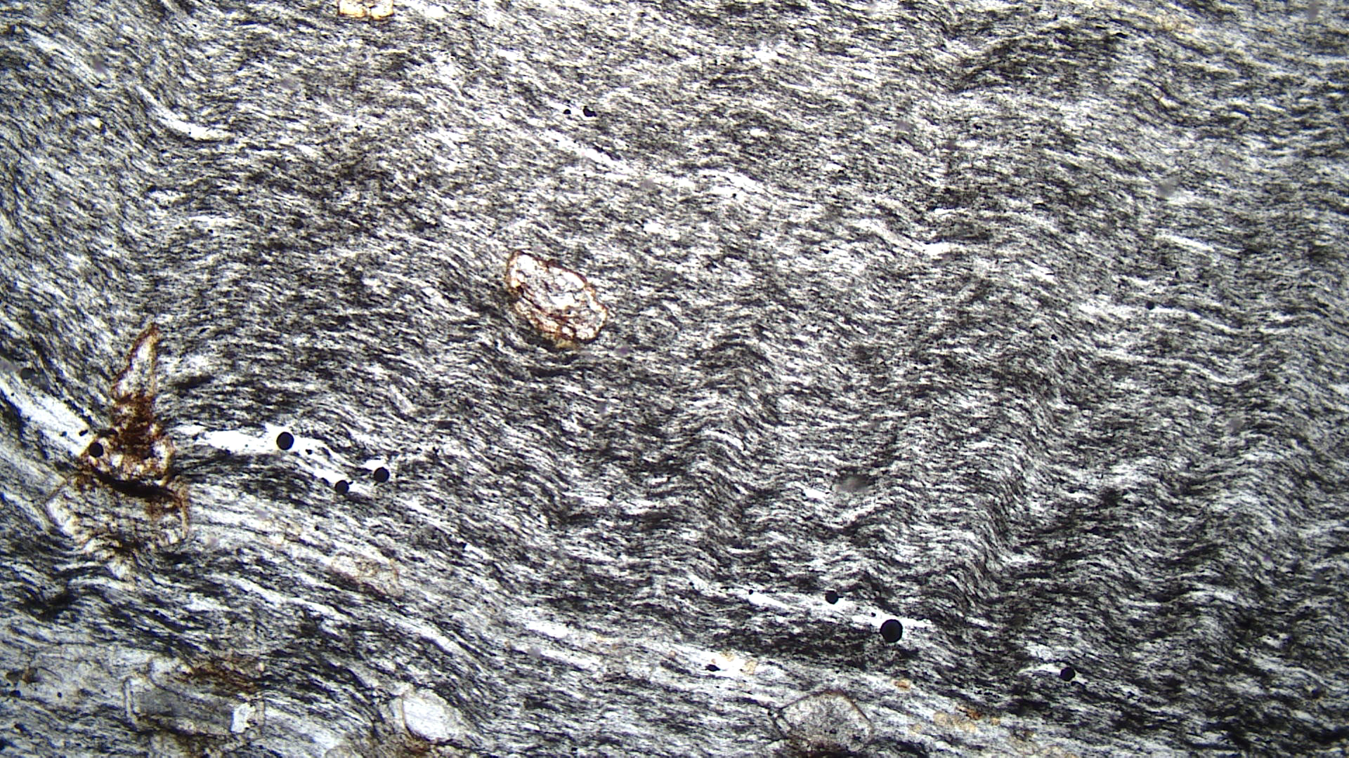
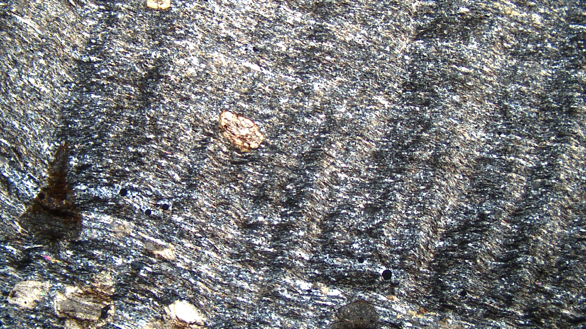
Greenstone is a general term we use to describe fine-grained unfoliated metamorphosed basalts. These rocks get their green color from epidote, chlorite, or actinolite. This greenstone, that comes from just west of Ely, Minnesota, is a composed mostly of actinolite and plagioclase. These minerals, however, cannot be distinguished in the hand-sample view.
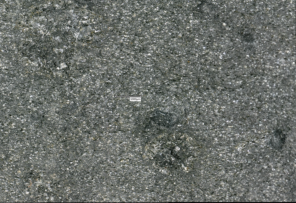
The plane-polars thin-section view contains elongate green actinolite and white plagioclase crystals. In crossed polars, the actinolite displays 2nd- and 3rd-order colors and plagioclase 1st-order colors. A few plagioclase grains exhibit twinning but, in general, the fine grain size makes twinning hard to see.
▪Greenschist
|
|
|
|

| ⇒Link to 3D rotatable image of a greenschist |
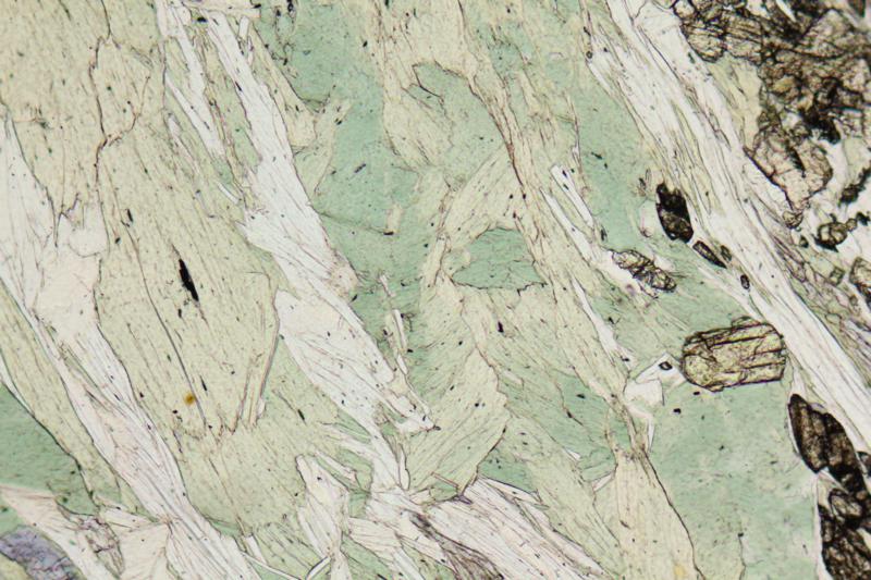
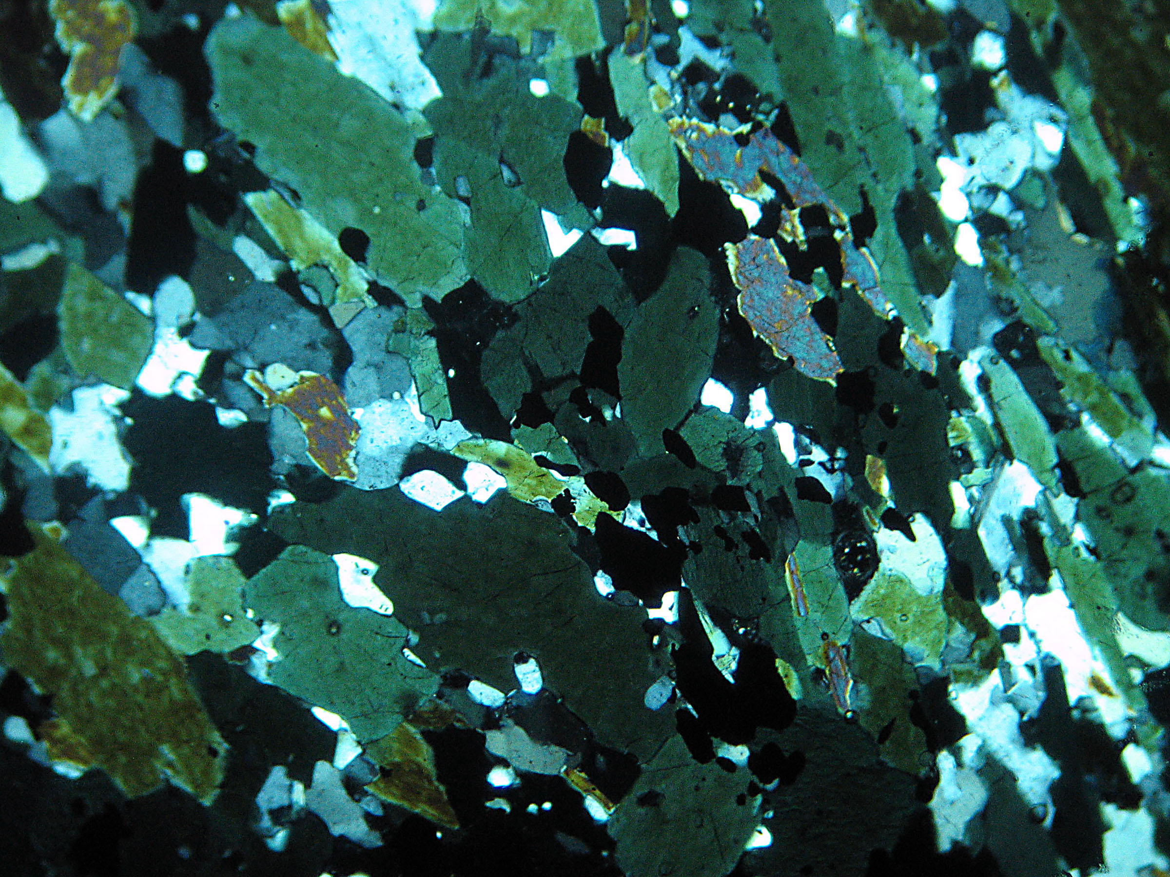
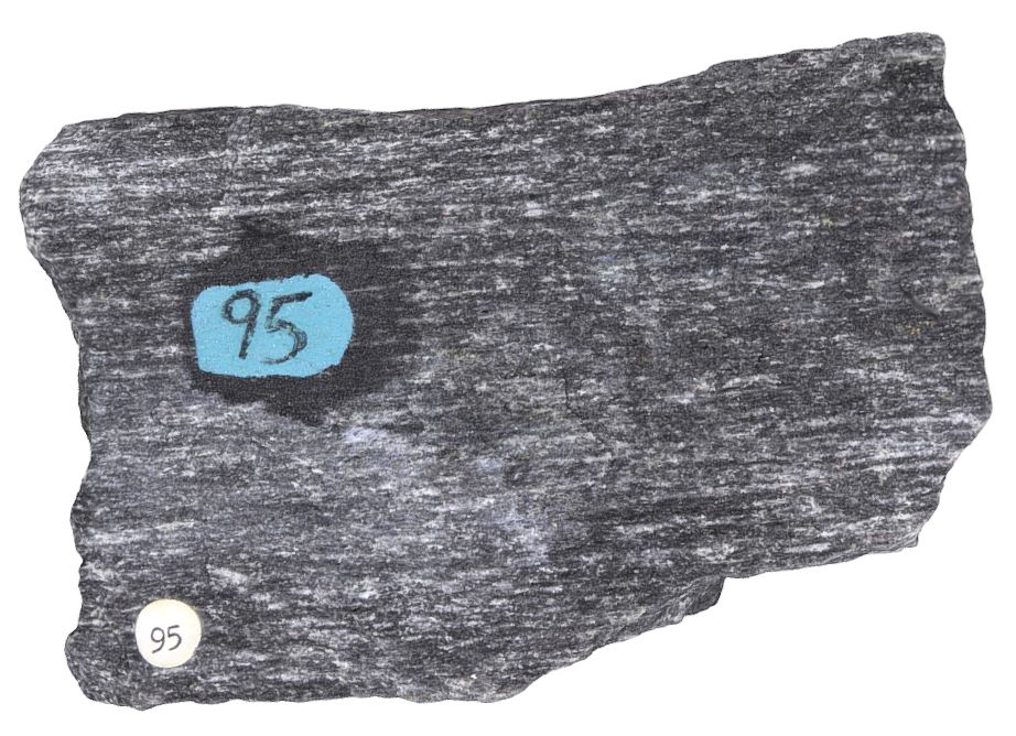
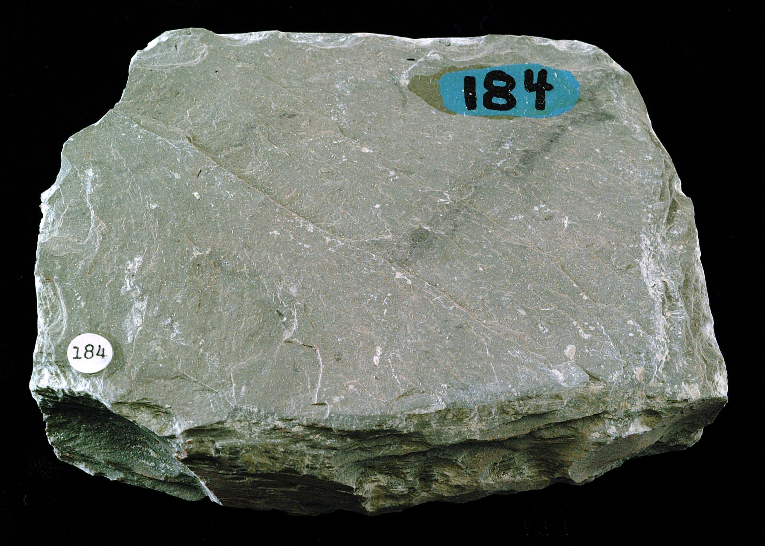
Greenschists are fine- to medium-grained foliated metamorphic rocks dominated by chlorite, actinolite or epidote. Na-plagioclase, quartz, muscovite, and calcite are sometimes present. The hand specimen in the photo on the left above is a greenschist from Michigan’s Upper Peninsula. The rock is made predominately of chlorite but is too fine-grained to see individual crystals.
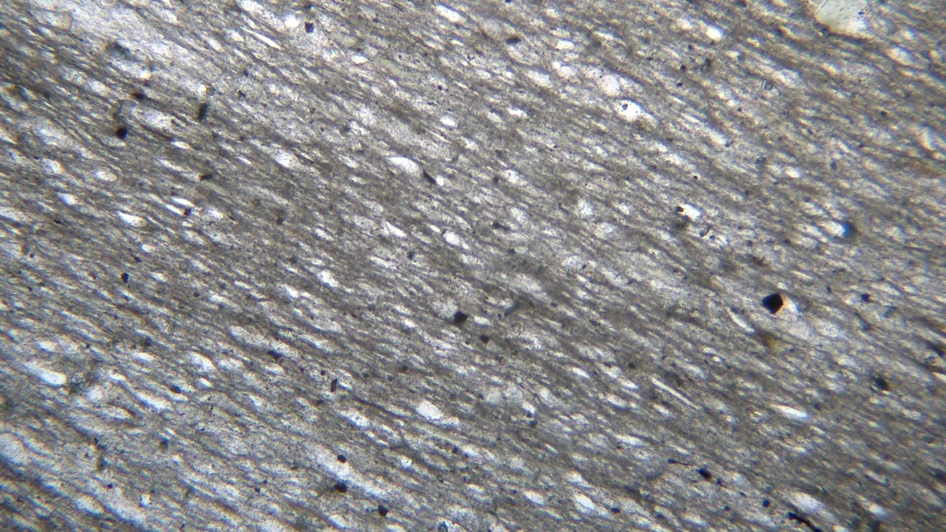
The thin-section views are of a different greenschist. The PP view shows chlorite in various shades of green, clear muscovite, and (near the right side) high-relief epidote. In crossed polars, the chlorite displays anomalous gray-green interference colors and the muscovite has up to 3rd-order colors. The epidote has very high-order colors that, in some grains, vary like an archery target from grain rim to center.
▪Actinolite Schist
|
|
|
|


| ⇒Link to 3D rotatable image of an actinolite-talc schist |
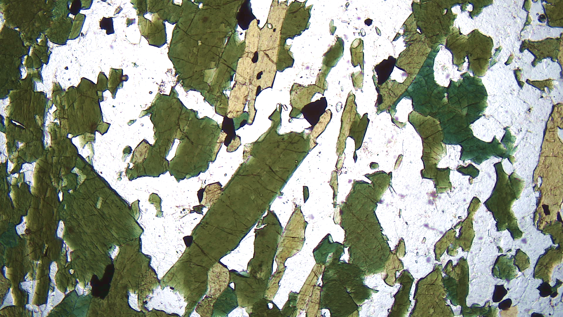
The photos above show a schist from Chester, Vermont, that contains conspicuous needles of green actinolite. Pleochroic green actinolite dominates the PP thin-section view. Clear quartz and opaque magnetite are also present. In the XP view, the deep color of the actinolite partially masks its interference colors, making them somewhat anomalous. A few of the amphibole grains have amphibole’s classic 60o-120o cleavage angles.
▪Hornblende Schist
|
|
|
|
| ⇒Link to 3D rotatable image of this amphibolite |
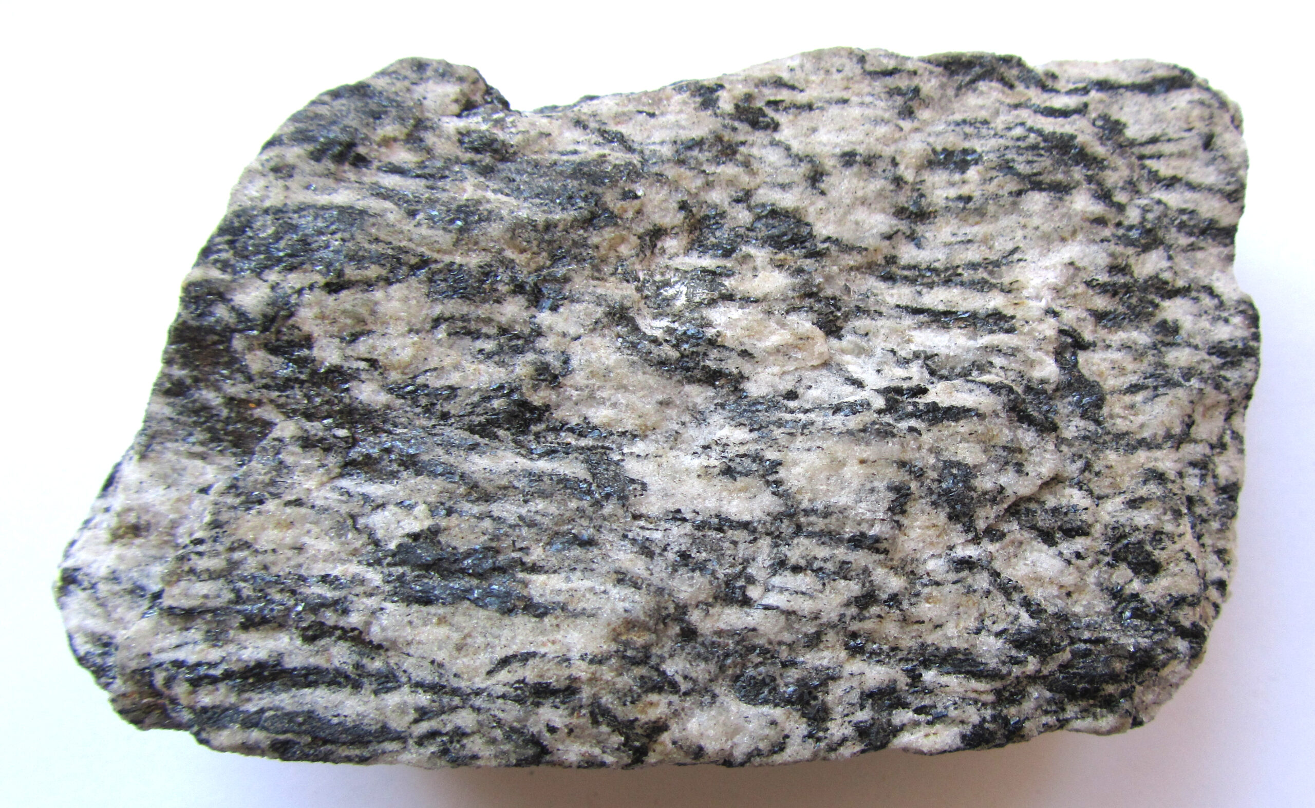
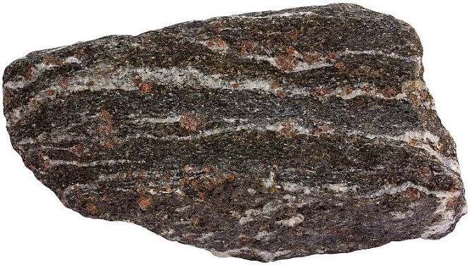
The hornblende schist in Figure 16.160 is from Mitchell County, North Carolina. The medium-sized black hornblende needles are easily seen in the hand specimen. The other major minerals are quartz and plagioclase but they are hard to tell apart. The rock also contains abundant epidote that cannot be seen in hand specimen. The many thin elongate layers in this rock suggest it was highly stressed while crystallizing.
In thin section, hornblende in this sample is pleochroic green to brown and has up to high 2nd-order interference colors. Quartz and minor plagioclase are clear and have 1st-order white to black interference colors. A few small plagioclase crystals show twinning, but otherwise distinguishing plagioclase from quartz is difficult without being able to rotate the thin section. High-relief epidote, present in patches in some parts of the sample, displays very high-order colors. Magnetite is also present.
▪Hornblende Schist
|
|
|
|

This hornblende schist comes from Gouverneur, New York. In hand specimen, especially when viewed with a hand lens, sparkly hornblende crystals appear to alternate with light-colored plagioclase crystals.
In thin section, the hornblende is pleochroic in shades of brown to green. It has up to high 2nd-order interference colors. Plagioclase and (minor) quartz are clear and have white to gray interference colors. Magnetite is also present, and a single grain of calcite can be seen on the bottom edge near the left side.
▪Amphibolite
|
|
|
|

This very fine-grained amphibolite comes from near Soudan, Minnesota. In hand specimen it appears dark colored and homogeneous. A hand lens reveals, however, that there are both light colored and dark colored mineral grains, although they cannot be identified with certainty.
In thin section, brown pleochroic hornblende and clear plagioclase are easily distinguished in XP light. The amphibole displays classic 60o-120o cleavage angles. Crossing the polars reveals that the plagioclase is twinned and has 1st-order white to black colors. The hornblende has up to 2nd-order interference colors. Minor magnetite is also present.
▪Hornblende Gneiss
|
|
|
|

| ⇒Link to 3D rotatable image of this hornblende gneiss |
This hornblende gneiss is from Clintonville, New York. The hand specimen shows moderate (gneissic) foliation due to parallel alignment of hornblende crystals and plagioclase-rich layers.
In thin section, hornblende appears pleochroic in browns and greens. The only other major mineral is plagioclase – it is quite altered and so has a rough grainy texture under PP light. High-relief titanite, and minor biotite and magnetite are also present. Under crossed polars, the hornblende displays up to 2nd-order blue and red interference colors. Plagioclase appears moth-eaten and has been substantially replaced by sericite. Abundant quartz and minor magnetite are also present.
▪Augite Gneiss
|
|
|
|

| ⇒Link to 3D rotatable image of this gneiss |
This augite gneiss is from Lowell, Idaho. The hand specimen contains cm-long tabs of augite floating in a sea of quartz and plagioclase. The rock contains substantially more quartz than plagioclase.
Augite crystals have light-green color in the PP thin-section view. Quartz and plagioclase are clear. Small grains of high-relief titanite are widespread as accessory minerals. In the XP view, quartz and feldspar have 1st-order interference colors, and augite has up to 3rd-order colors. The plagioclase crystals display fine polysynthetic twinning.
▪Garnet Amphibolite
|
|
|
|

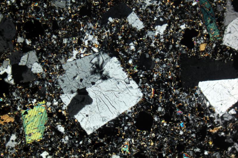
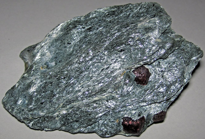
The hand specimen is a photo of a typical garnet amphibolite. It contains plagioclase (white), hornblende (black), and garnet porphyroblasts (red).
In the PP thin-section view, hornblende is green and pleochroic. A single large high-relief garnet is just right of the center of the photos (fractured with a pinkish color). The surrounding clear grains, aligned with the hornblende, are plagioclase with minor quartz.
In the crossed-polars view, the garnet is isotropic and the hornblende has up to 2nd-order, or low 3rd-order, interference colors. The plagioclase has 1st-order white to black interference colors with indistinct albite twinning.
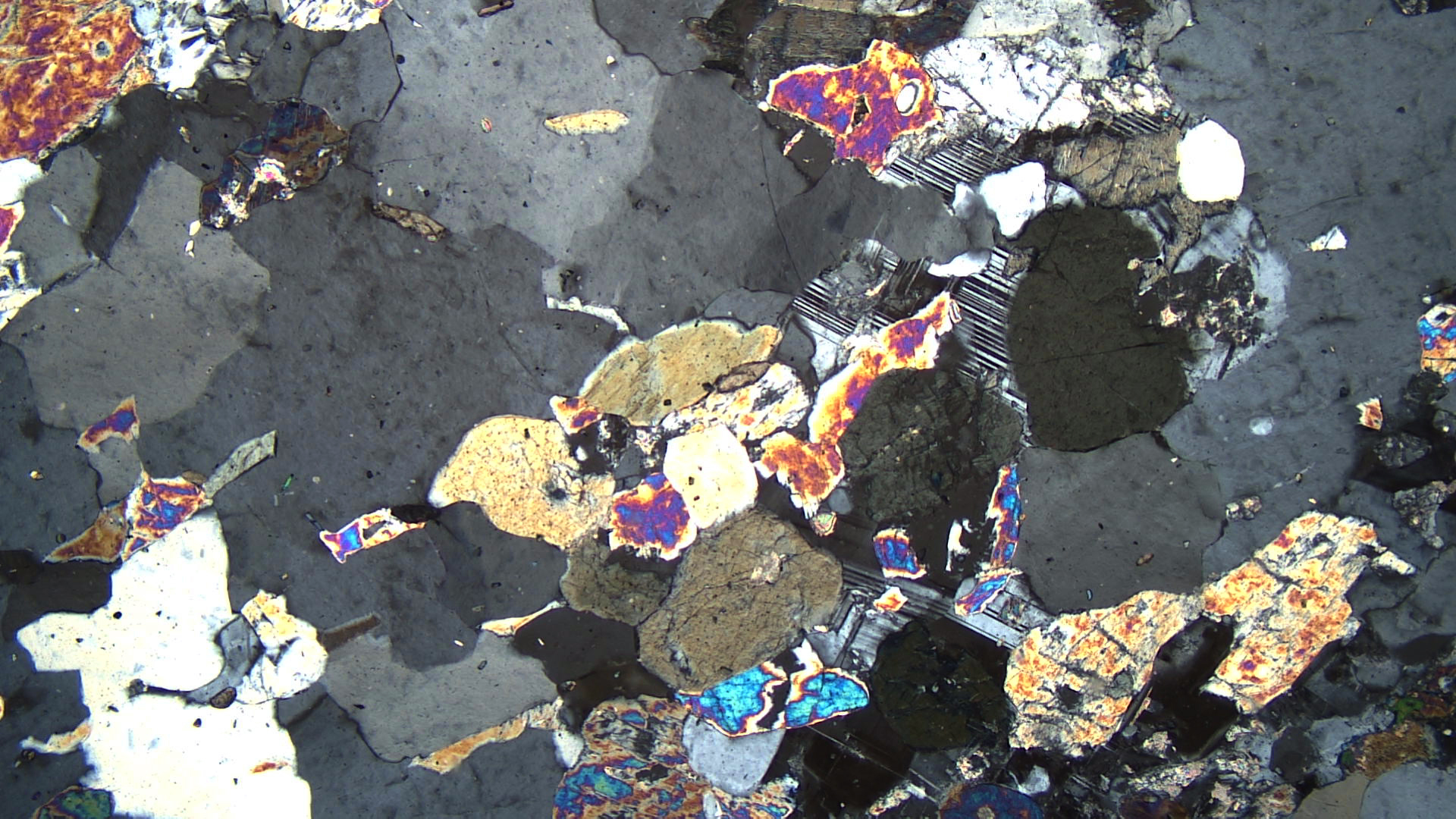
▪Garnet Granulite
|
|
|
|
The cobble hand specimen seen above is a mafic granulite from the Ivrea Zone, Italy. Minerals easily seen in the photo include red garnet, black clinopyroxene, and white plagioclase. Orthopyroxene is also present but not distinguishable.
The thin-section views are of a different mafic granulite. This one is from Hartmannsdorf, Germany. The photo using plane-polarized light shows clinopyroxene (pleochroic green), orthopyroxene (pleochroic pink to green), and plagioclase (colorless). The colors of the orthopyroxene and garnet are quite similar. There is one large garnet crystal, just right and up from the center of the photograph (pinkish). Abundant opaque magnetite is scattered throughout.
In crossed polars the garnet is isotropic and the plagioclase has 1st-order colors with most grains showing twinning. The clinopyroxene grains have up to low 3rd-order interference colors, and the orthopyroxene grains has high 1st- to 2nd-order interference colors.
16.3.3 High-Pressure Rocks
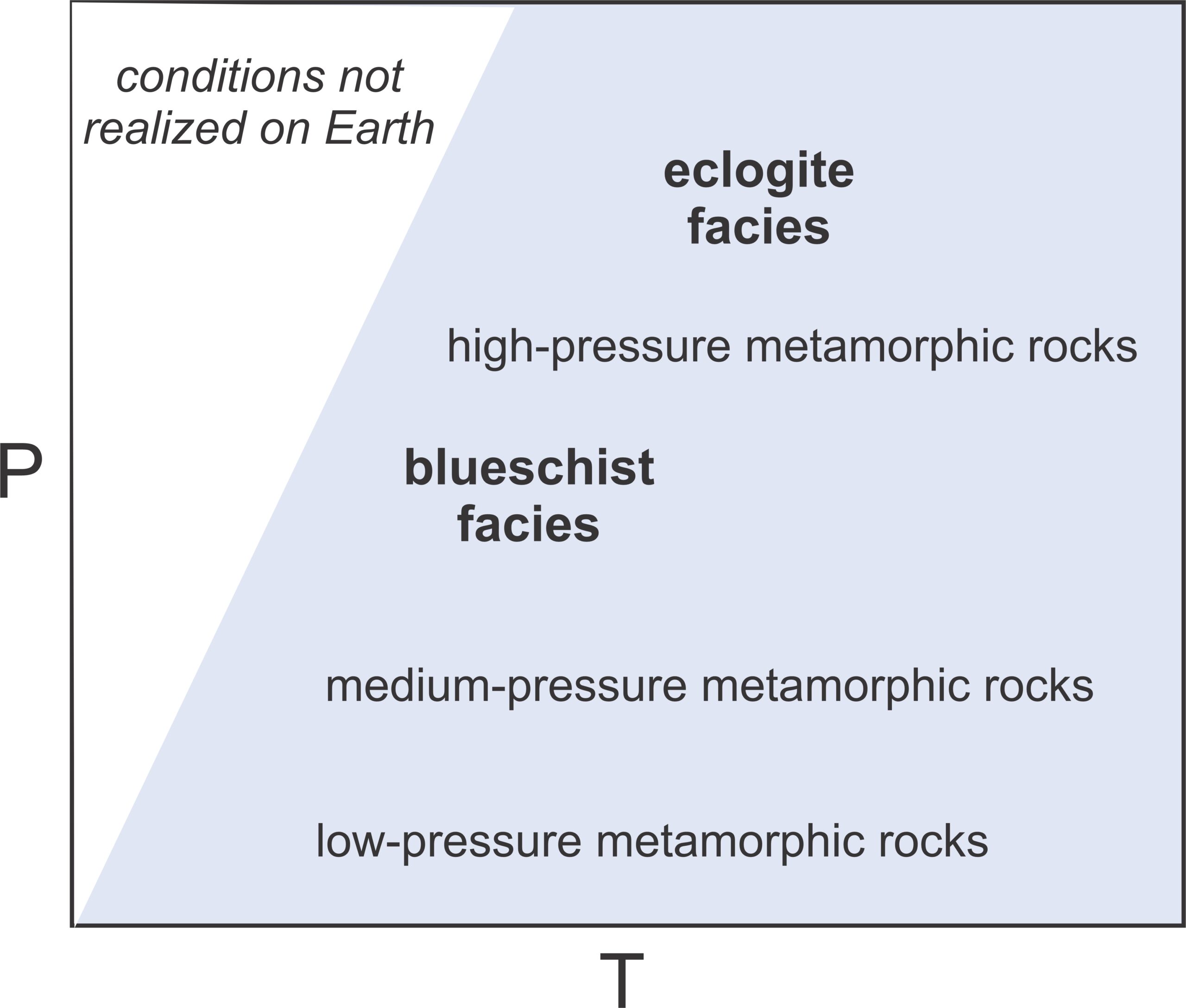
16.181 High-pressure metamorphic facies
As seen in Figure 16.181, high-pressure metamorphic rocks include primarily rocks of the blueschist and eclogite facies. Most blueschist- and eclogite-facies rocks are mafic. Blueschists (including glaucophane schists) are generally associated with subduction zones. Eclogites, too, may occasionally be associated with subduction zone metamorphism. They are also found in ophiolite complexes and as xenoliths carried to the surface from great depth.
▪Glaucophane Schist
|
|
|
|


| ⇒Link to 3D rotatable image of this glaucophane schist |
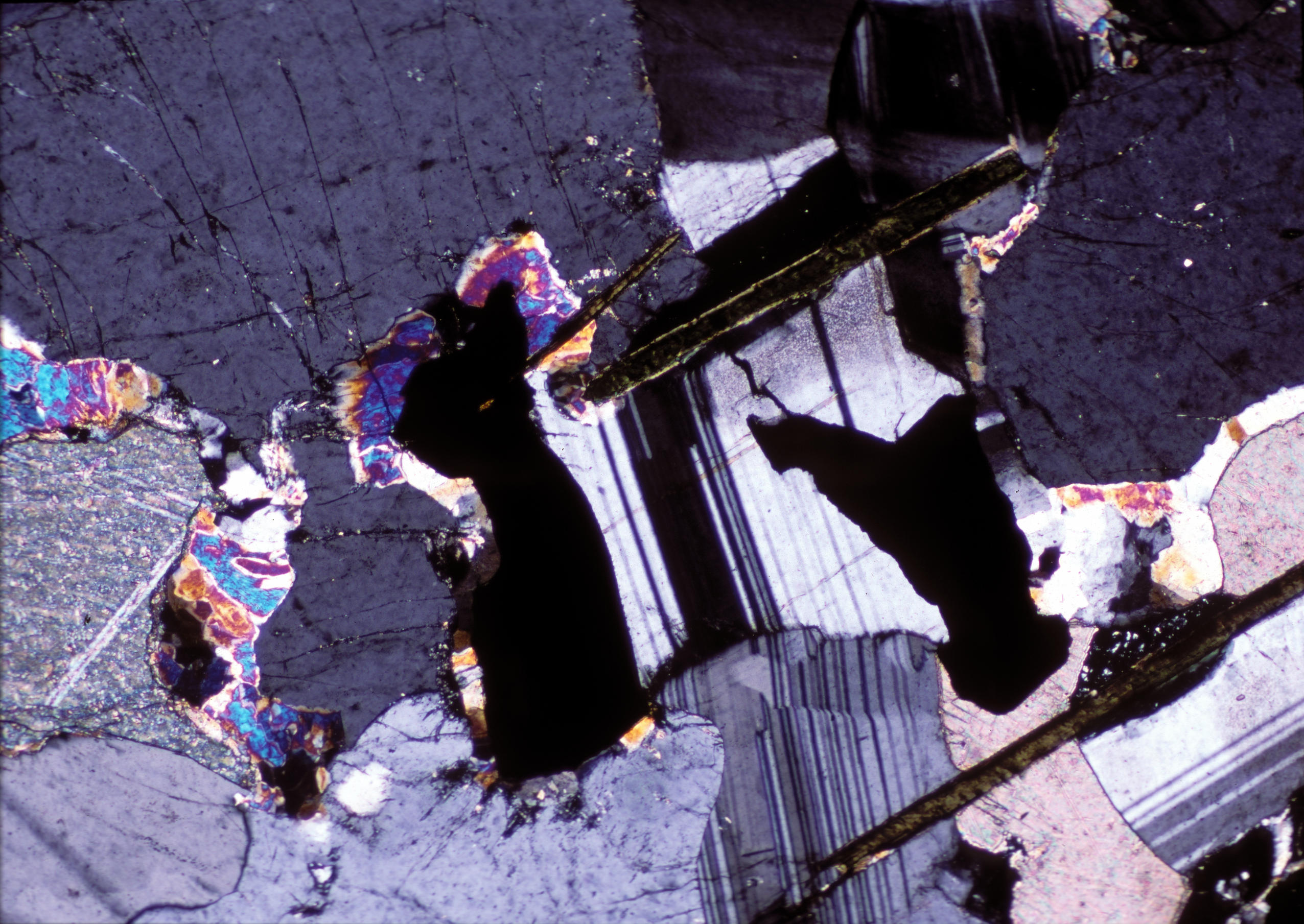
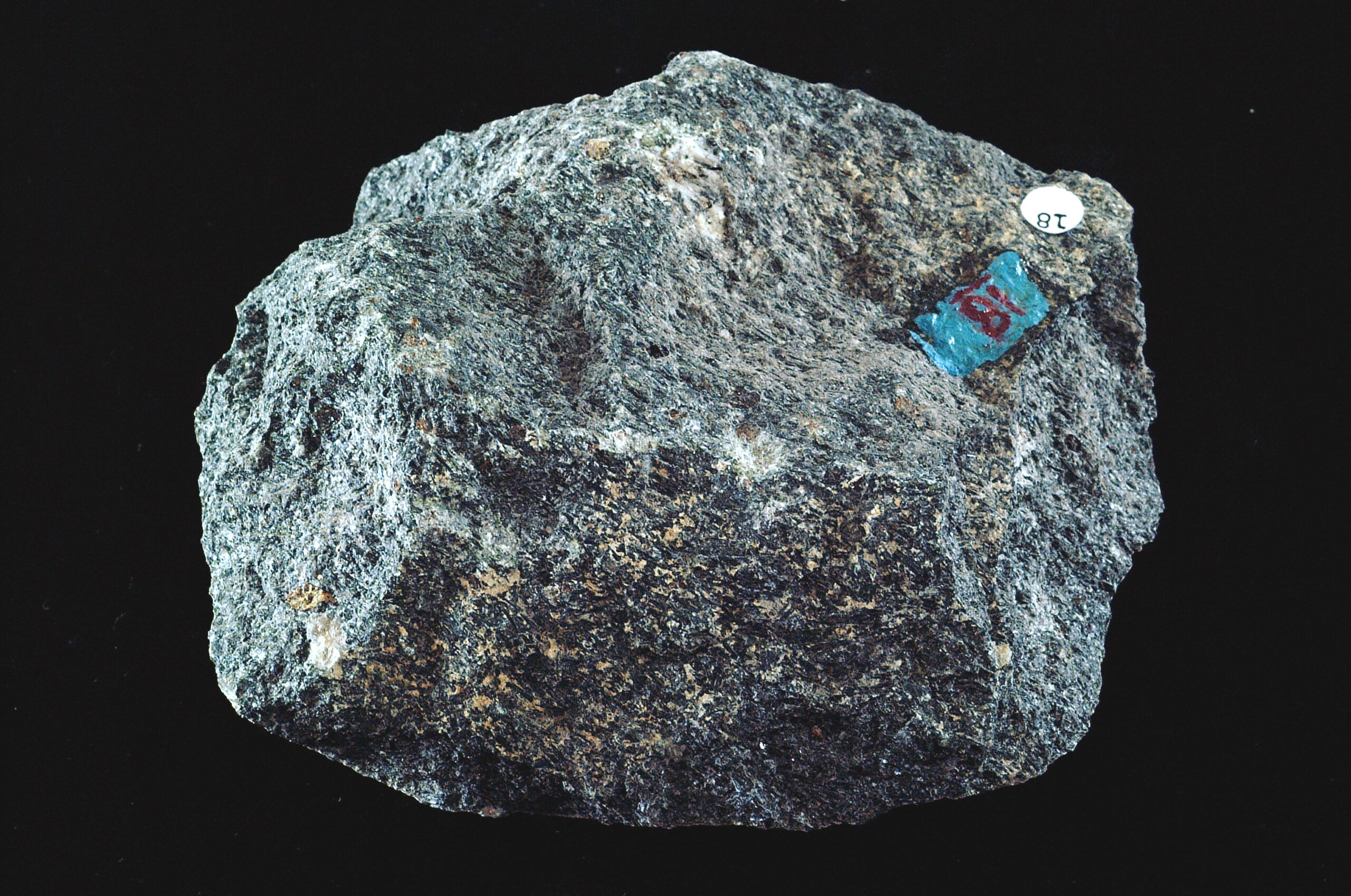
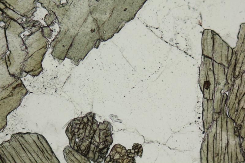
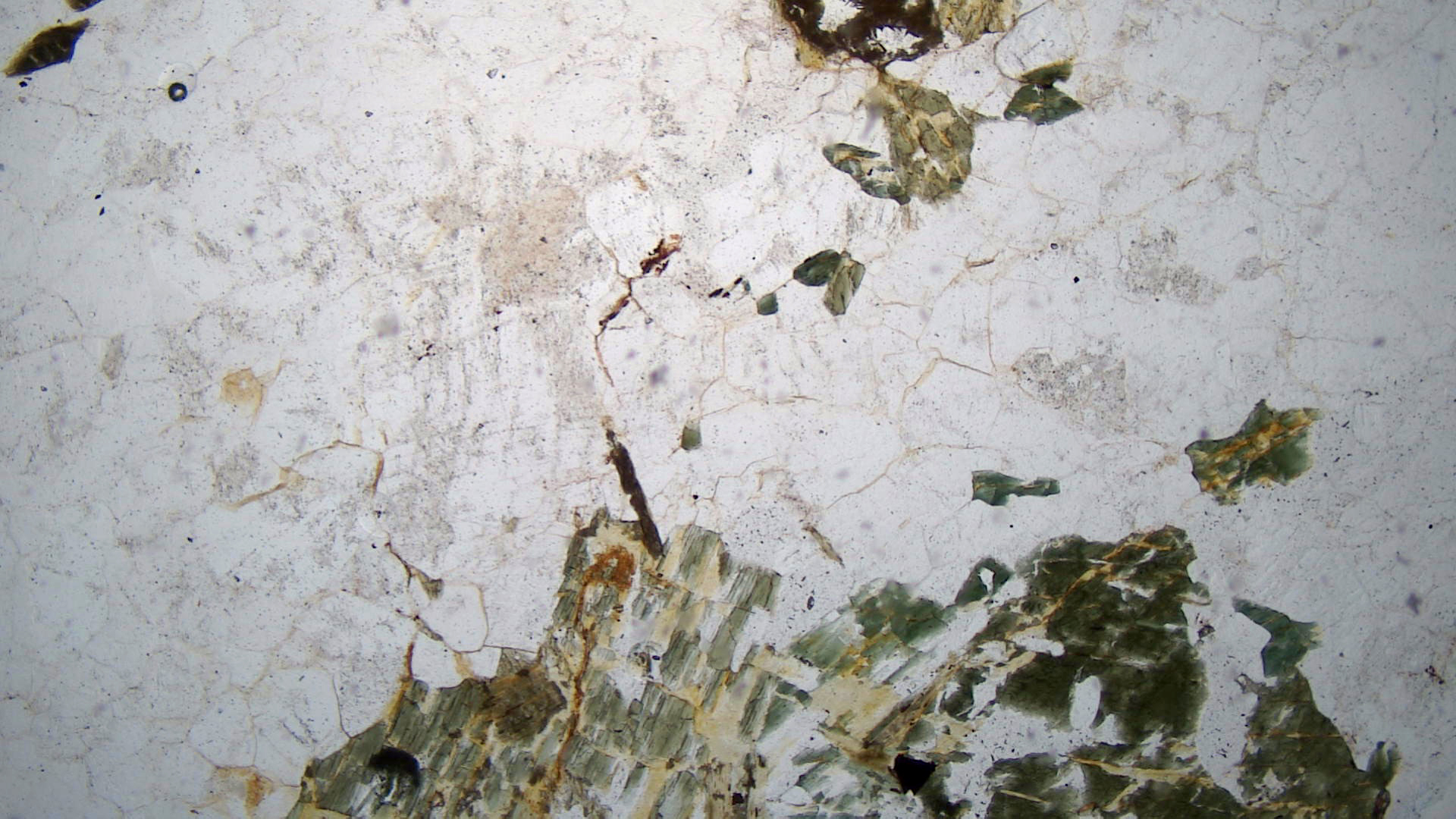
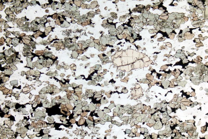
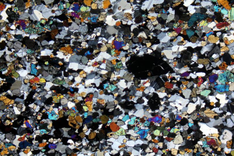
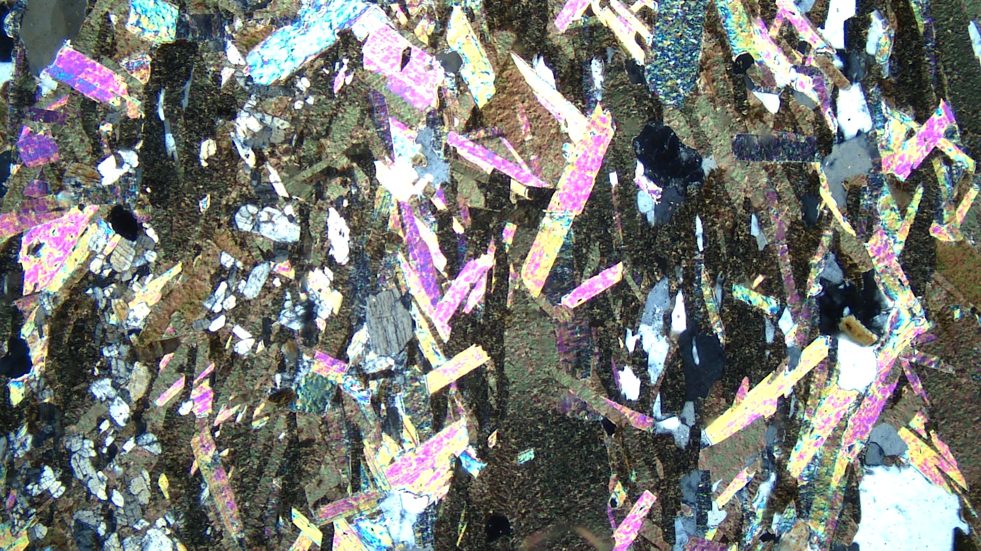
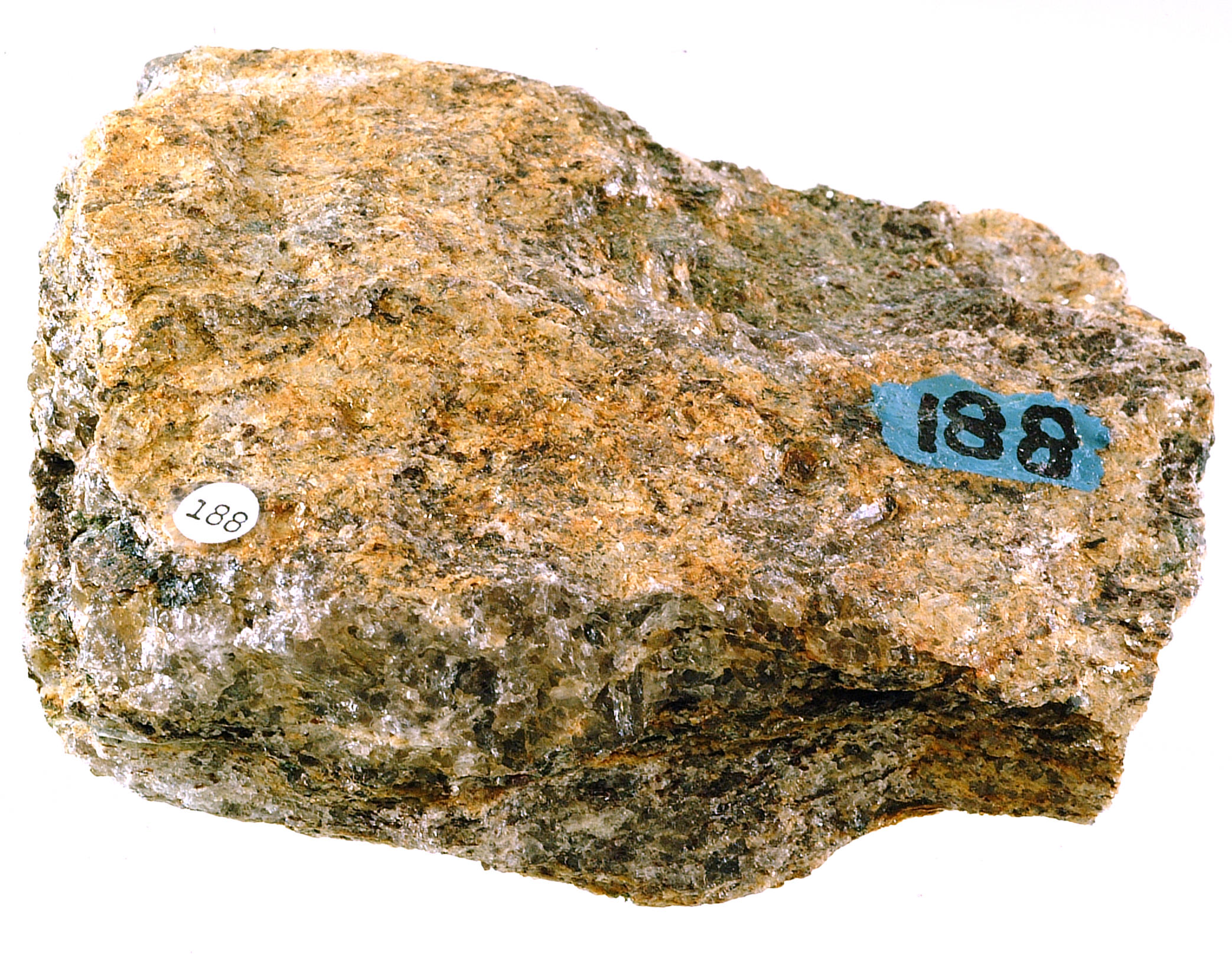
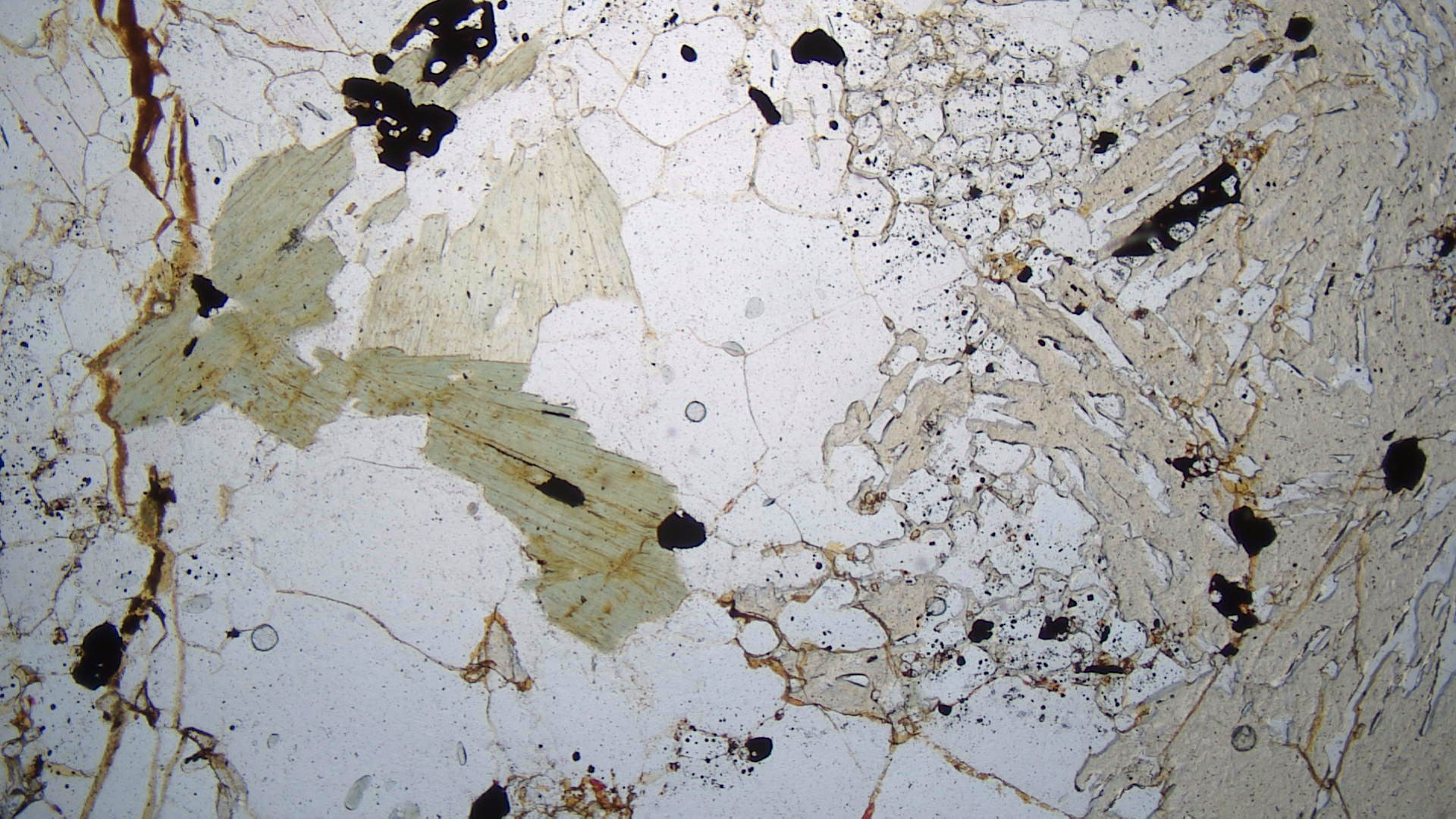
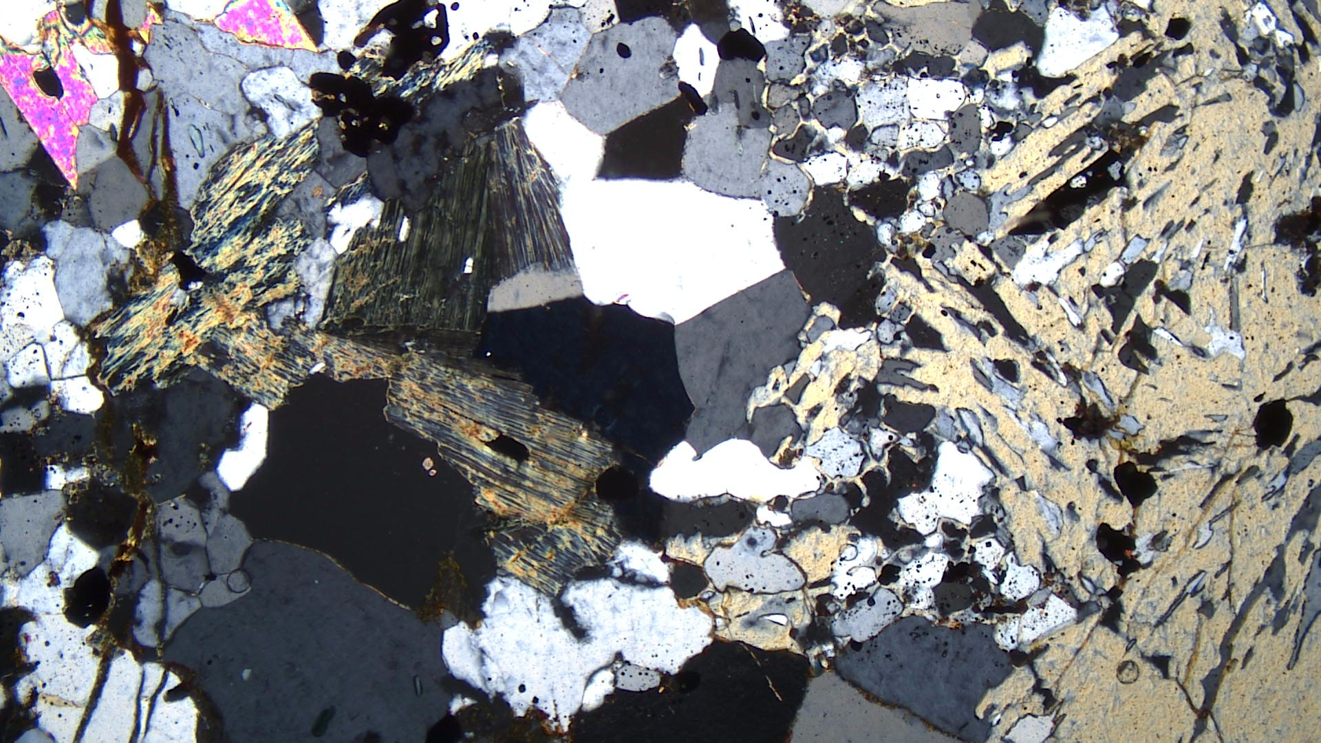
This glaucophane schist (hand specimen and thin-section views) comes from Sonoma County, California. Glaucophane schists are also call blueschists. This specimen contains primarily blue-gray glaucophane. Minor epidote give the hand specimen a slightly green hue in some places. In plane-polarized light, the thin section reveals mostly blades of light-blue glaucophane (labeled Glauc). The light-green mineral with relatively high relief in the PP view is epidote (labeled Epi). Several small grains of titanite are present and stand out because of their high relief.
Note that, in the XP view, glaucophane’s interference colors are high order in some grains and appear anomalous in others. This is because the color of the mineral has added to the normal upper 1st-order interference colors in some grains. Epidote displays 3rd-order or higher patchy interference colors.
▪Blueschist
|
|
|
|
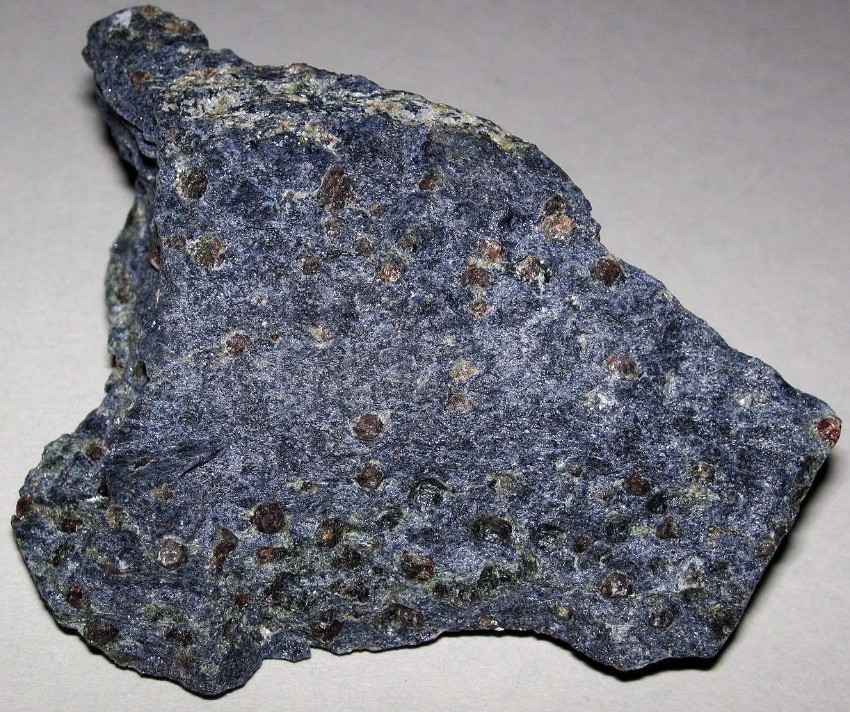
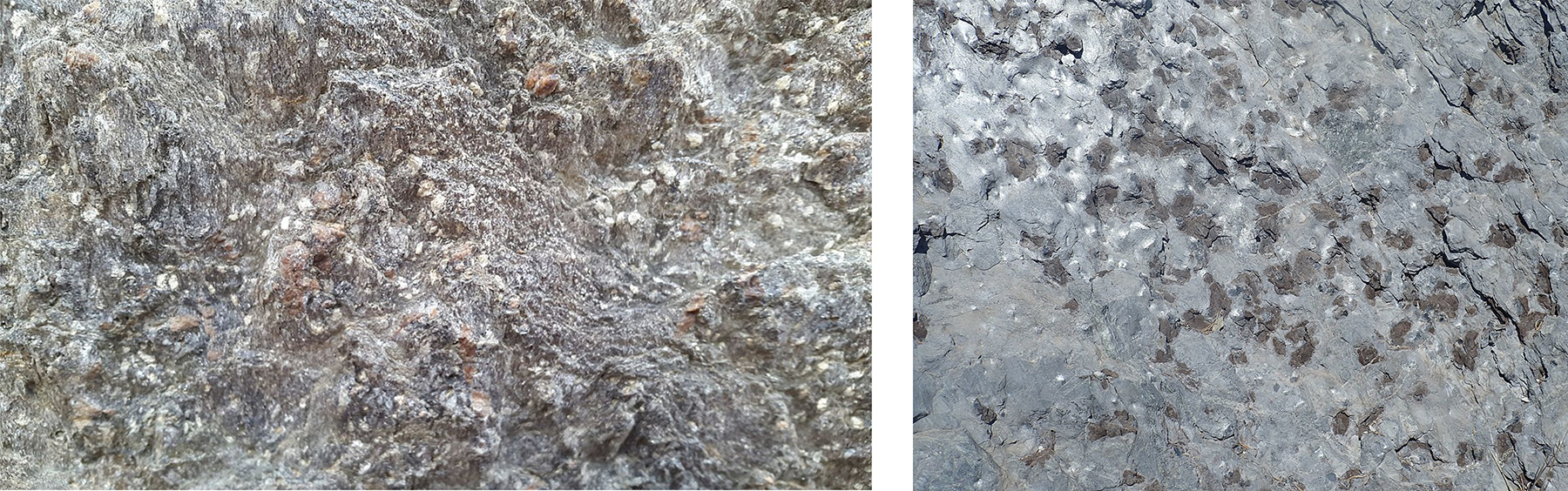
The blueschist hand specimen in the photo above comes from a famous locality near Cazadero, California. The outcrop seen on the right is a photo of a blueschist knocker in Sonoma County, California. Knocker is the term used by California geologists to describe out-of-context blocks of erosion-resistant rocks surrounded by softer materials. This knocker is near the top of a hill that overlooks San Francisco Bay.
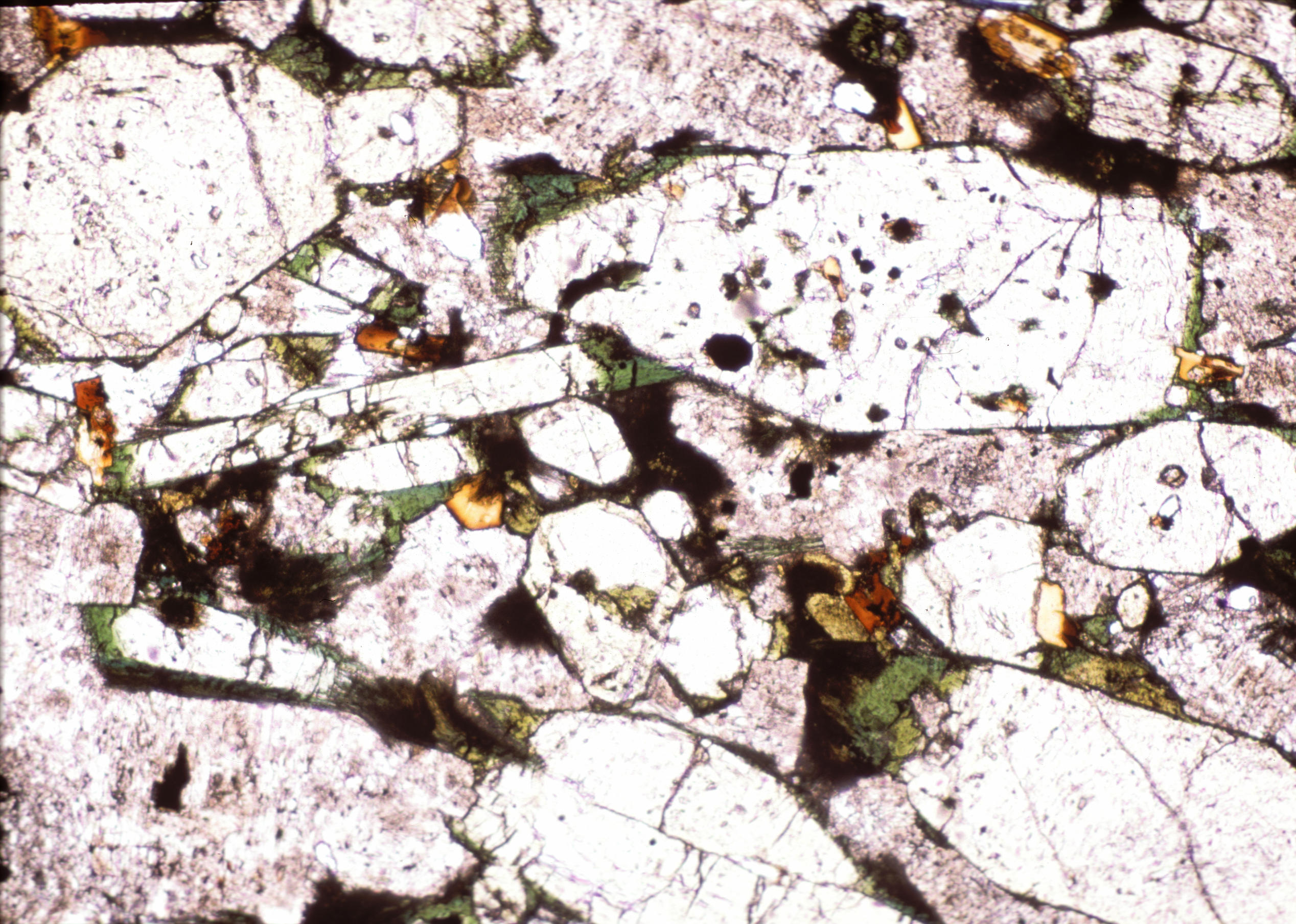
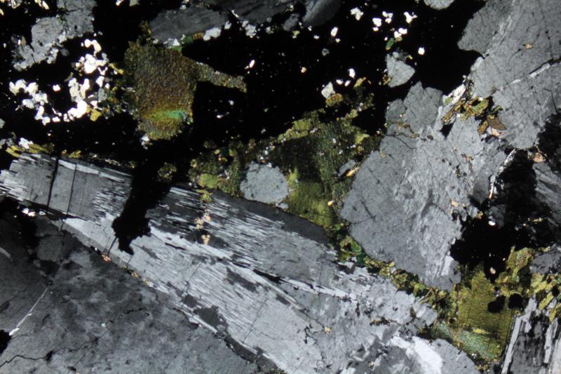
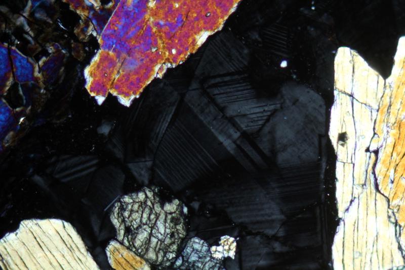
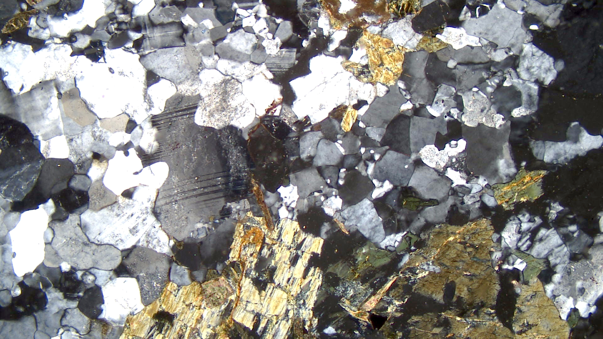
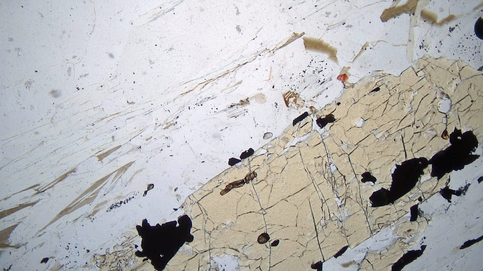
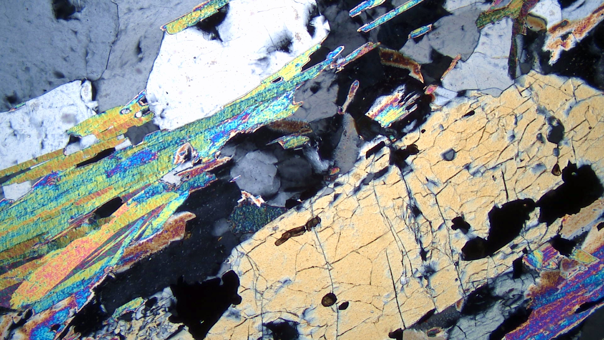
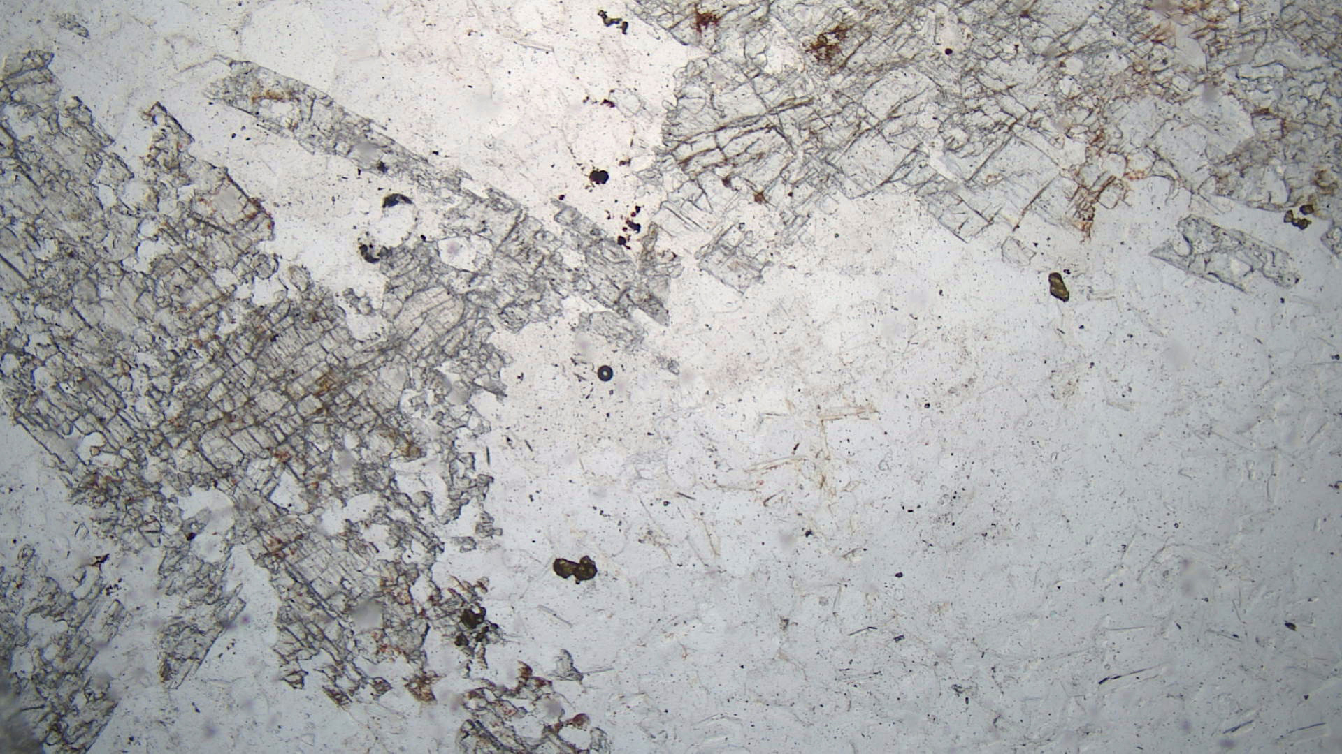
The blueschist seen in thin section is from a different place. It comes from Palos Hill on the island of Syros, Greece. In plane-polarized light, the thin section shows blue-violet glaucophane. Some of the glaucophane in this rock is in prismatic crystals with diamond-shaped cross sections showing cleavages at 60o from each other. Blue-violet cross sections of those crystals can be seen up and right from the center of this photo. Prisms lying parallel to the slide are lighter blue and appear as broad flat regions in the photo. A large grain of light green omphacite (a clinopyroxene) can be seen on the right side of the photo (PP view); smaller omphacite grains are also present. Most of the clear regions are white mica, probably phengite. Small high-relief grains are epidote and titanite, and opaque magnetite is present in small amounts.
In crossed polars, the white mica has high-order interference colors. Glaucophane has low 1st-order to upper 2nd-order interference colors (the diamond-shaped cross sections). Omphacite has low birefringence and interference colors are no greater than 1st-order yellow.
▪Eclogite
|
|
|
|


The hand specimen in the photo above is an eclogite from Norway’s Western Gneiss Region. It contains easily seen large red garnets, green omphacite (pyroxene), and white quartz. The thin-section views are of a different eclogite. Pink garnet and green omphacite are easily seen in the PP view. The clear mineral is quartz. The very deep brown mineral (it is almost black in the PP view) is rutile.
In crossed polars, the garnet is isotropic, the omphacite has up to high 1st-order interference colors, and the quartz has low 1st-order colors. Rutile has very high birefringence, but its interference colors are masked by the strong dark color of the mineral.
16.3.4 Metapelites and Metaquartzites
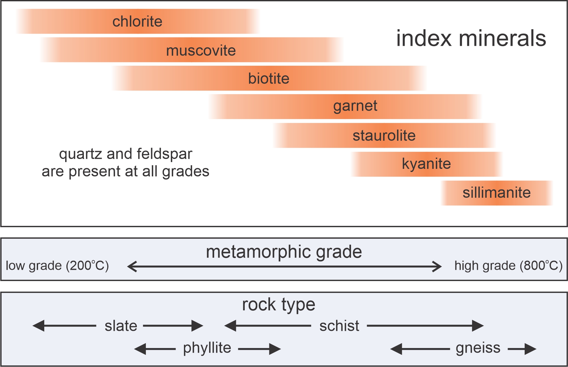
16.192 Index minerals and different kinds of rocks
Metapelites are metamorphosed clay-rich rocks. Protoliths include both mudstones and shales. In the following discussion, we also look at some examples of metaquartzites that contain aluminous minerals, because these are the same minerals that characterize metapelites.
During regional metamorphism, pelites, perhaps more than any other composition rocks, undergo distinctive changes in both mineralogy and texture. With increasing grade, metamorphism produces a series of index minerals: chlorite, muscovite, biotite, garnet, staurolite, kyanite, and sillimanite. The exact minerals that develop vary somewhat with rock composition.
Although the index minerals depicted in Figure 16.192 are typical, at lower pressures, and during contact metamorphism, andalusite may replace kyanite and sillimanite, and cordierite may be present instead of garnet. And at high temperature, muscovite and biotite both become unstable and break down into anhydrous minerals or may melt. Orthopyroxene can develop in high-grade rocks that contain little water.
Besides gaining new metamorphic minerals, pelites also develop metamorphic fabrics. From low grade to high grade, metapelites become slates, phyllites, schists, and gneisses. Figure 16.192 shows the rough correlation of the different fabrics with the appearances of different index minerals. The rocks described below follow from low grade to high grade.
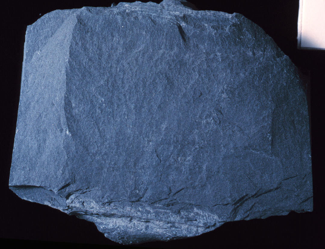
▪Gray Slate
|
|
|
|
| ⇒Link to 3D rotatable image of this gray slate |
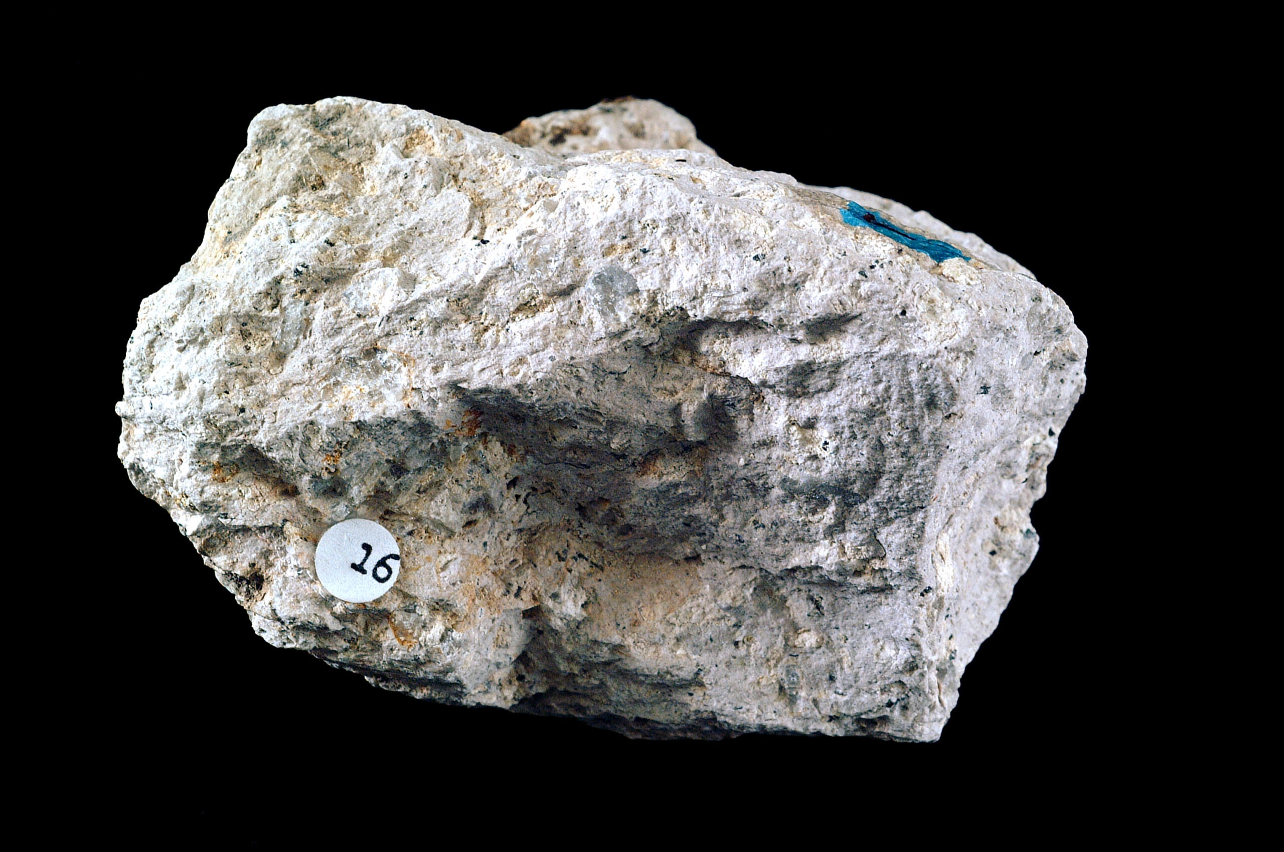
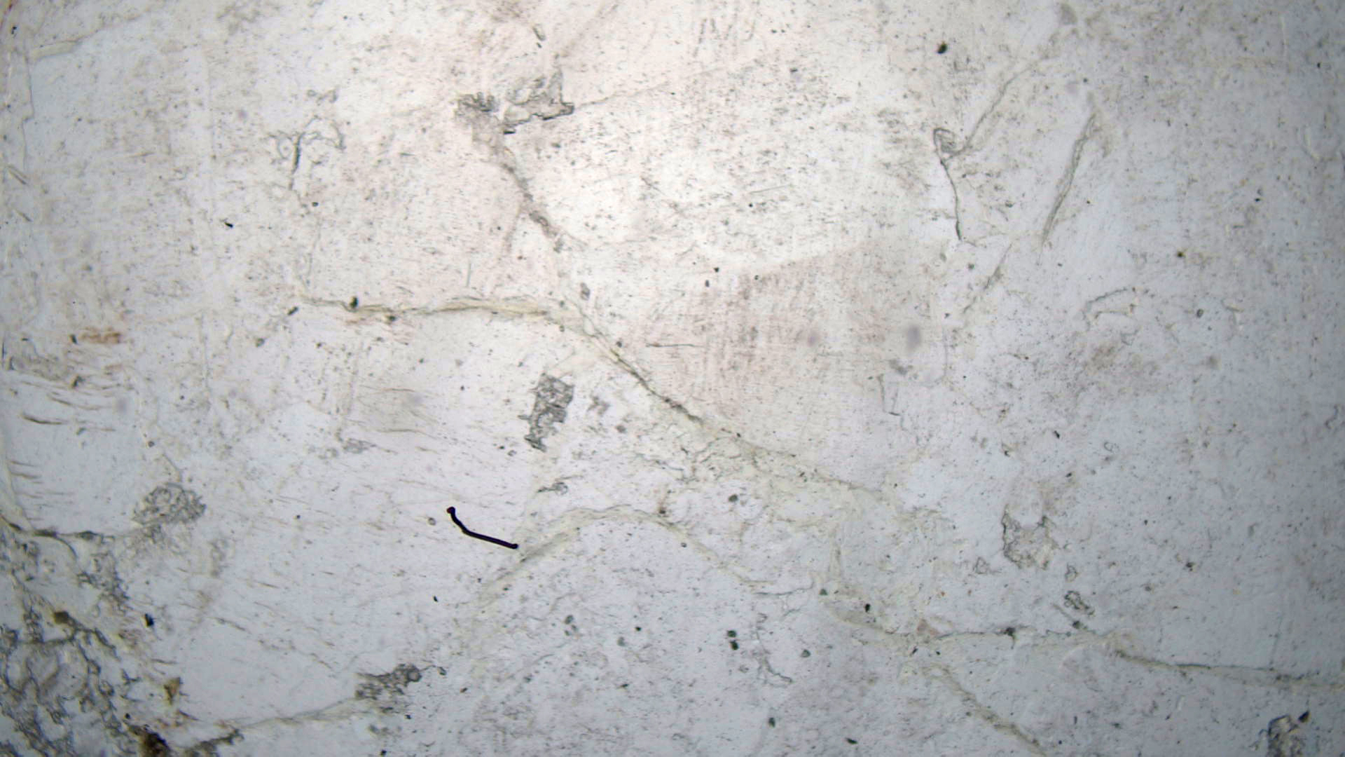
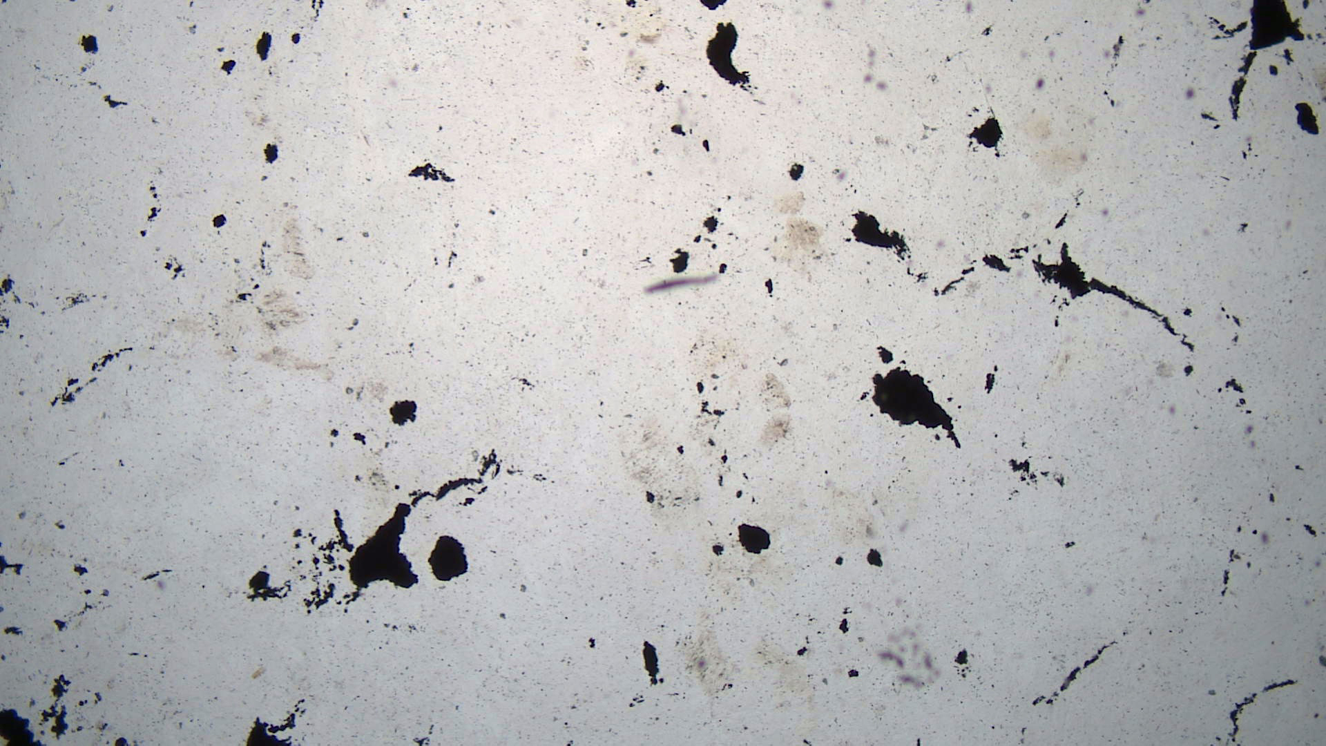
This is a sample of the Bangor slate, from near Stroudsburg, Pennsylvania. It is a very fine-grained rock with a well developed planar cleavage. The sample is so fine grained that identifying minerals in the hand specimen is impossible.
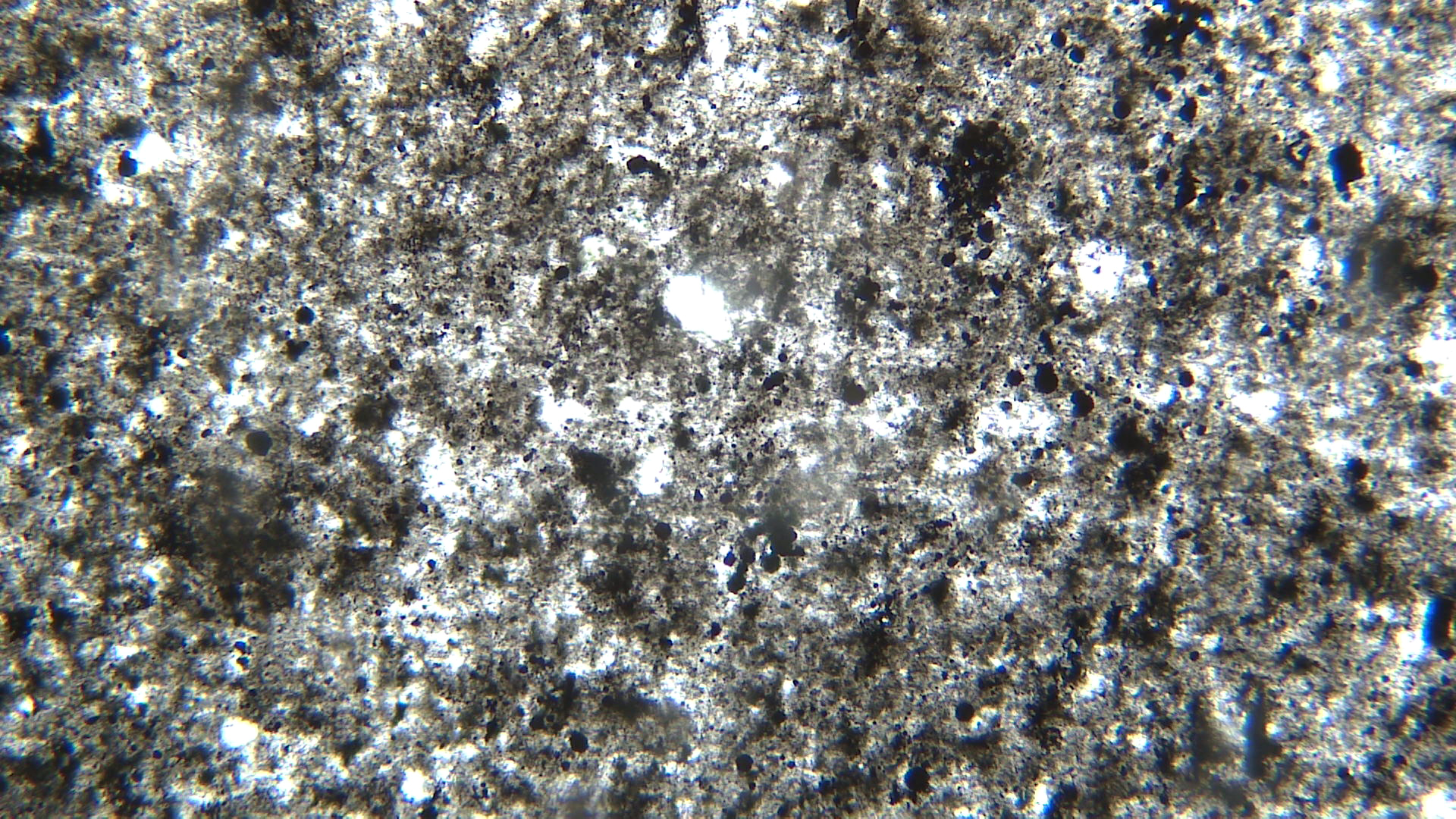
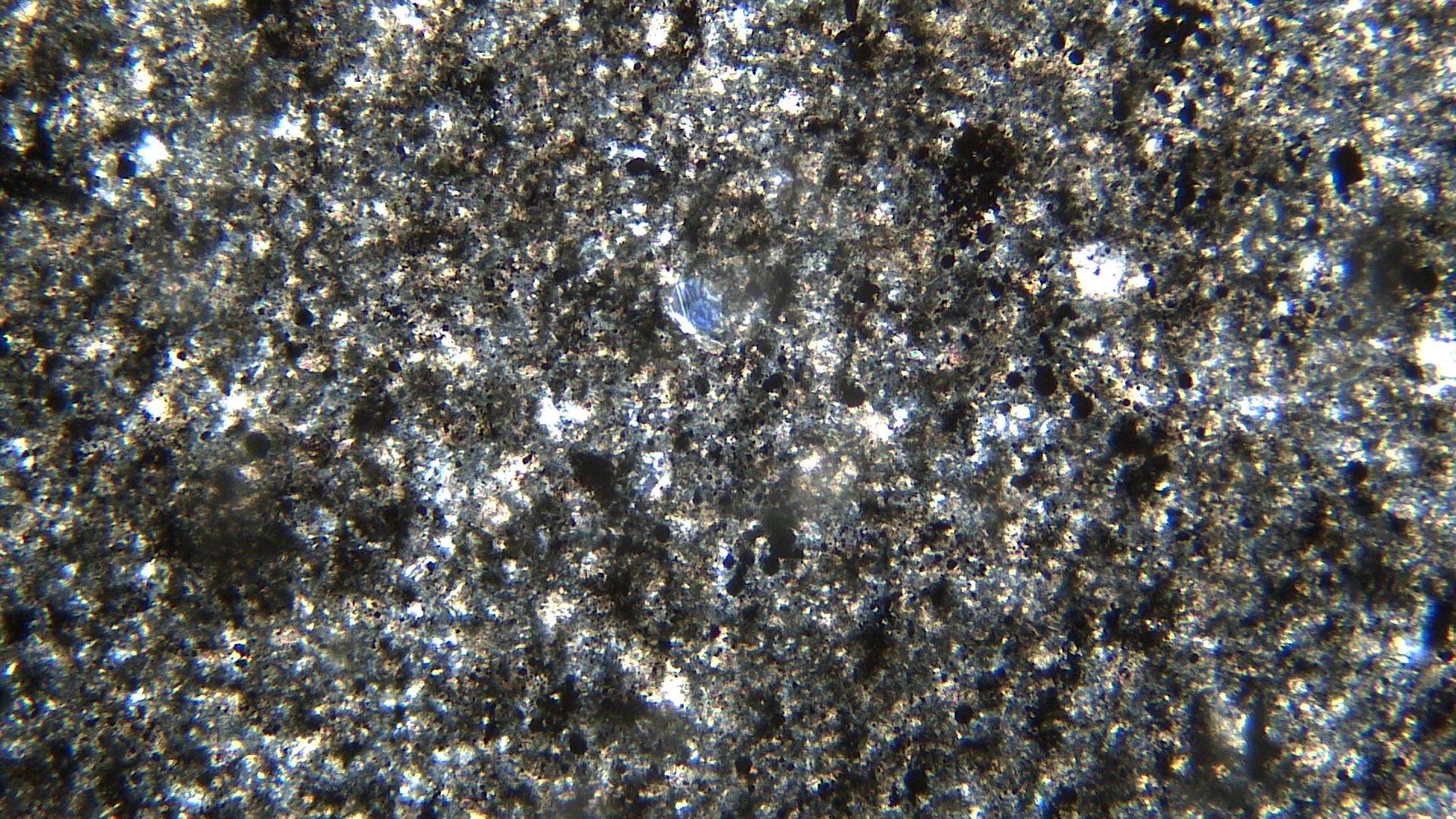
Due to fine grain-size, identifying minerals in the thin-section views is also difficult. The opaque minerals are graphite and magnetite. The clear minerals are mostly calcite and quartz. Small flakes of muscovite are present and display hints of 2nd- to 3rd-order interference colors in the XP view. Minor small flakes of chlorite are also present and have anomalous interference colors.
▪Andalusite Slate
|
|
|
|


| ⇒Link to 3D rotatable image of this andalusite slate |
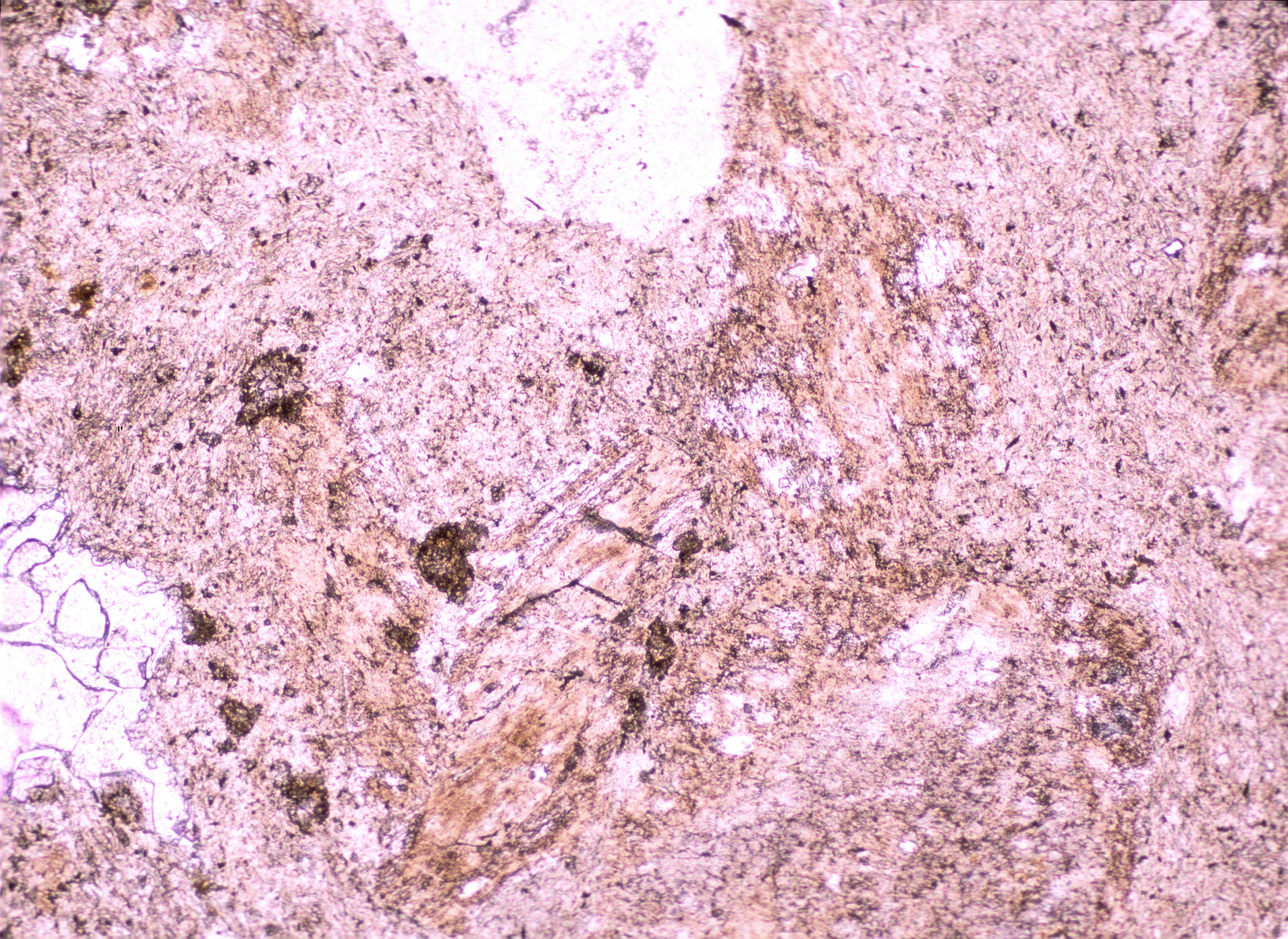
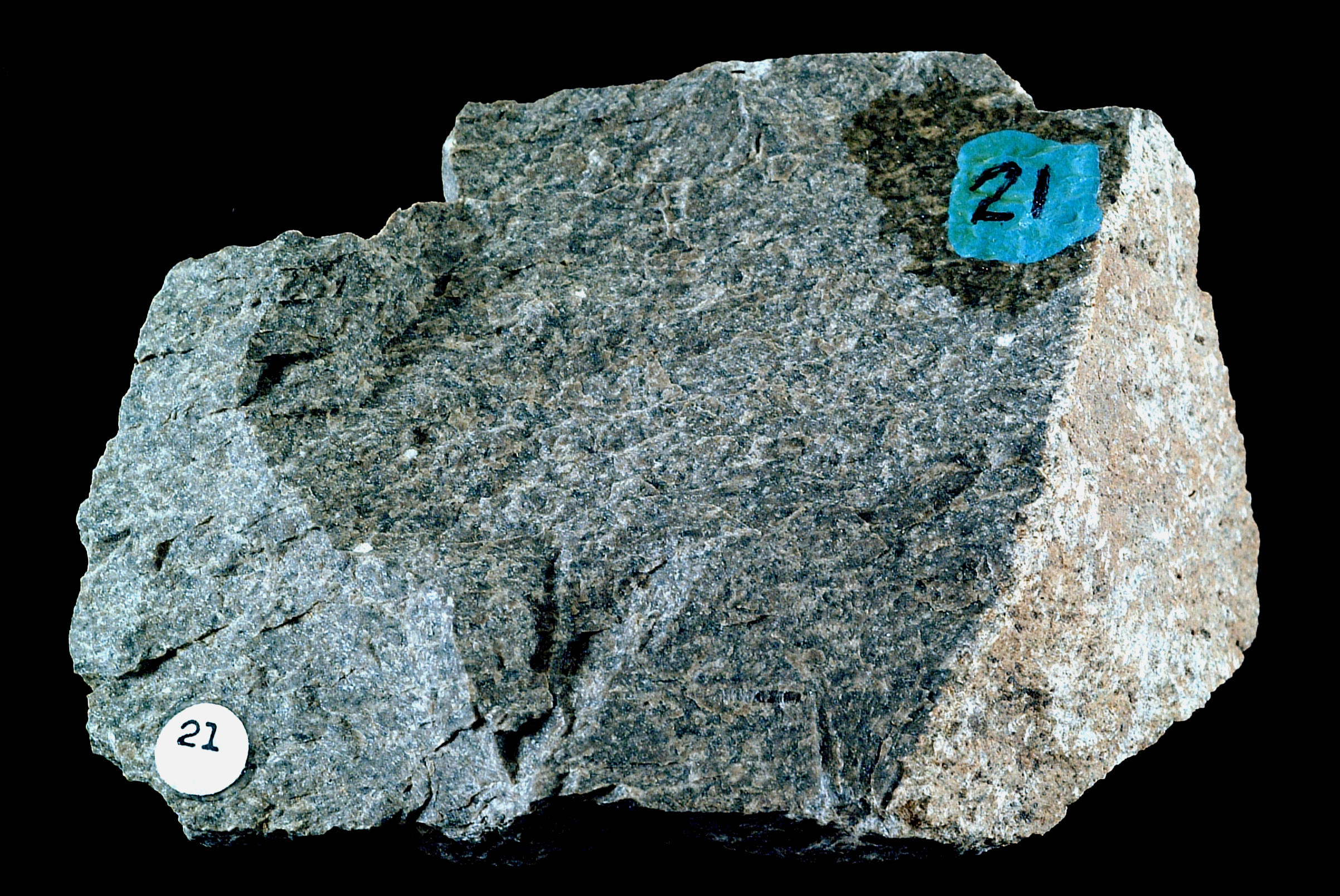
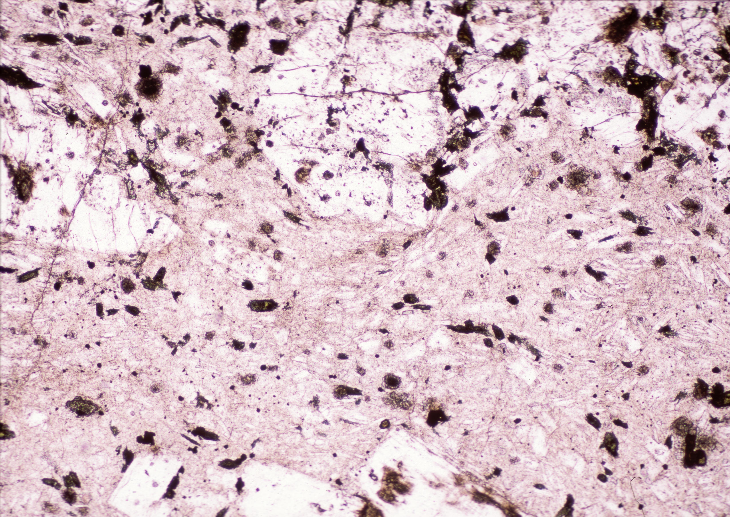
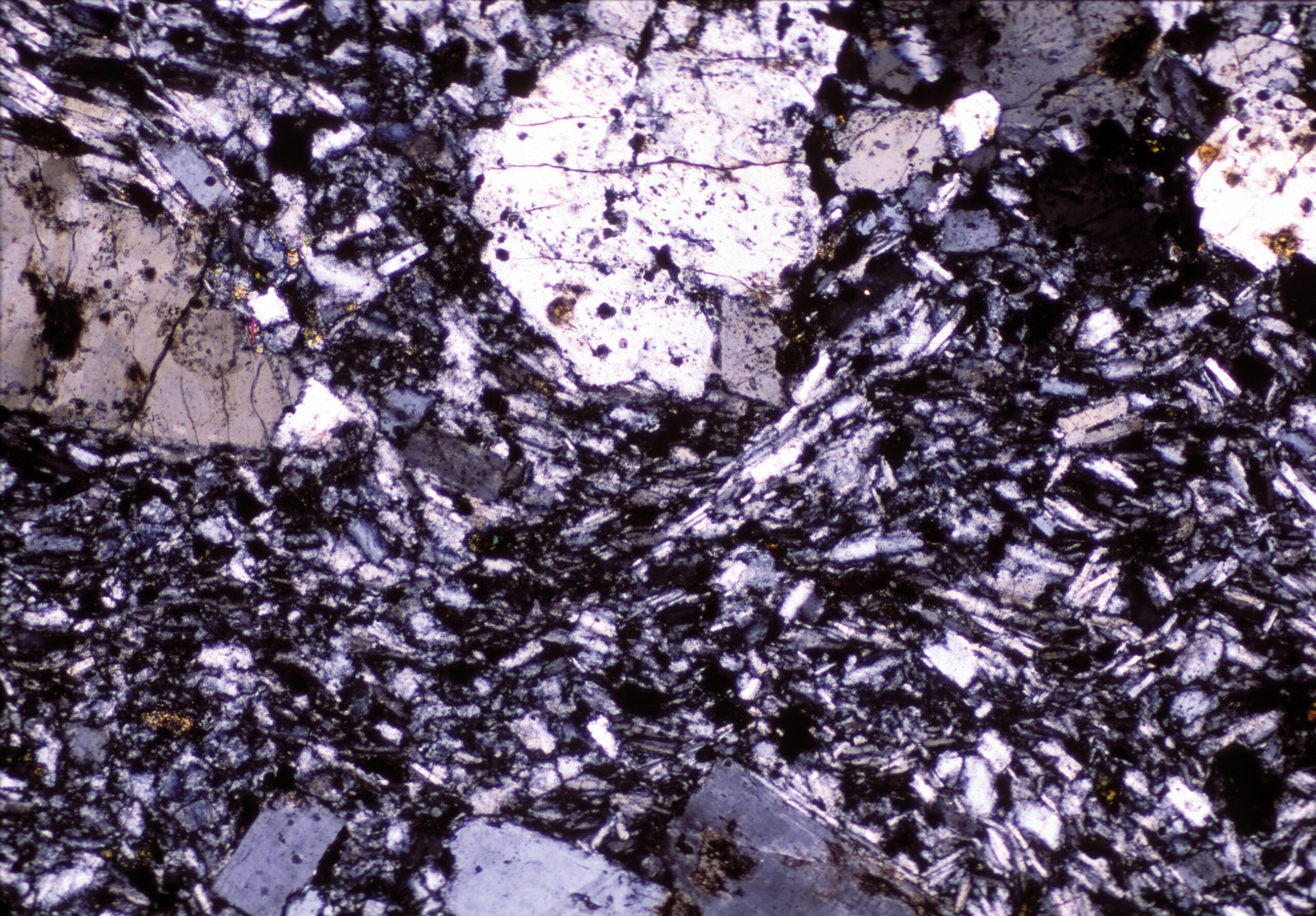
This dark-colored rock has a generally uniform color and fine-grained texture. However it contains small blocky andalusite crystals up to 1 or 2 mm across. They are hard to see in the hand specimen because they are about the same color as the finer-grained material that surrounds them. Figure 16.199 shows an enlarged view of the same rock and the andalusite crystals can just be seen. In higher-grade rocks, andalusite crystals may become quite large and have more distinct colors.
The PP and XP thin-section views of this sample (Figures 16.197 and 16.198, above) appear quite similar because almost none of the minerals present have high birefringence. One large andalusite crystal is on the upper left side of the thin-section; another shows partially on the upper right side. The large crystal displays vague concentric compositional zoning. None of the andalusite in this rock has the characteristic penetration twins that some andalusite displays. The fine groundmass is mostly a mix of quartz, graphite, and biotite. In the XP view, the small biotite crystals show brownish interference colors.
▪Green Slate
|
|
|
|
This green slate comes from Pawlet, Vermont. It is a very fine-grained rock that displays slaty cleavage when viewed parallel to its flat surfaces. It displays a slight hint of shiny reflectivity, so it is almost a phyllite.
The PP thin-section view exhibits the foliation quite clearly. Quartz grains (clear) are elongated and parallel. There are a few opaque mineral grains. The darker-colored gray whispy material that appears parallel to the foliation cannot be identified.
In crossed polars, quartz has its normal interference colors. Some small flakes of muscovite can be identified by their 2nd- to 3rd-order interference colors. The dark whispy material still cannot be identified.
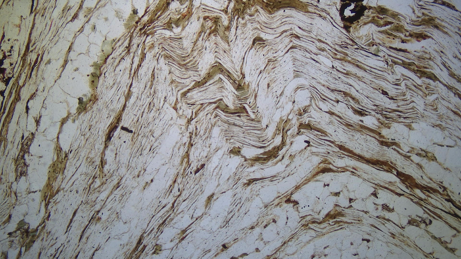
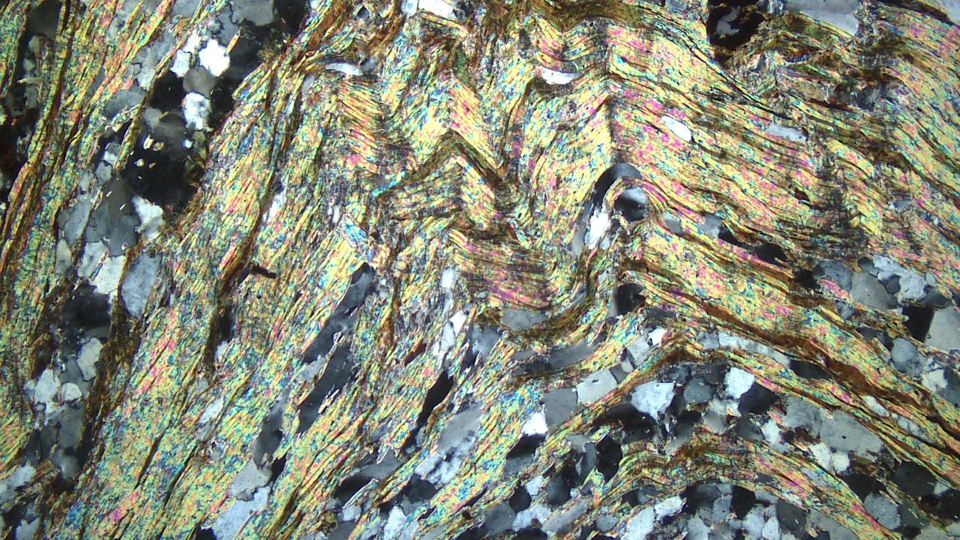
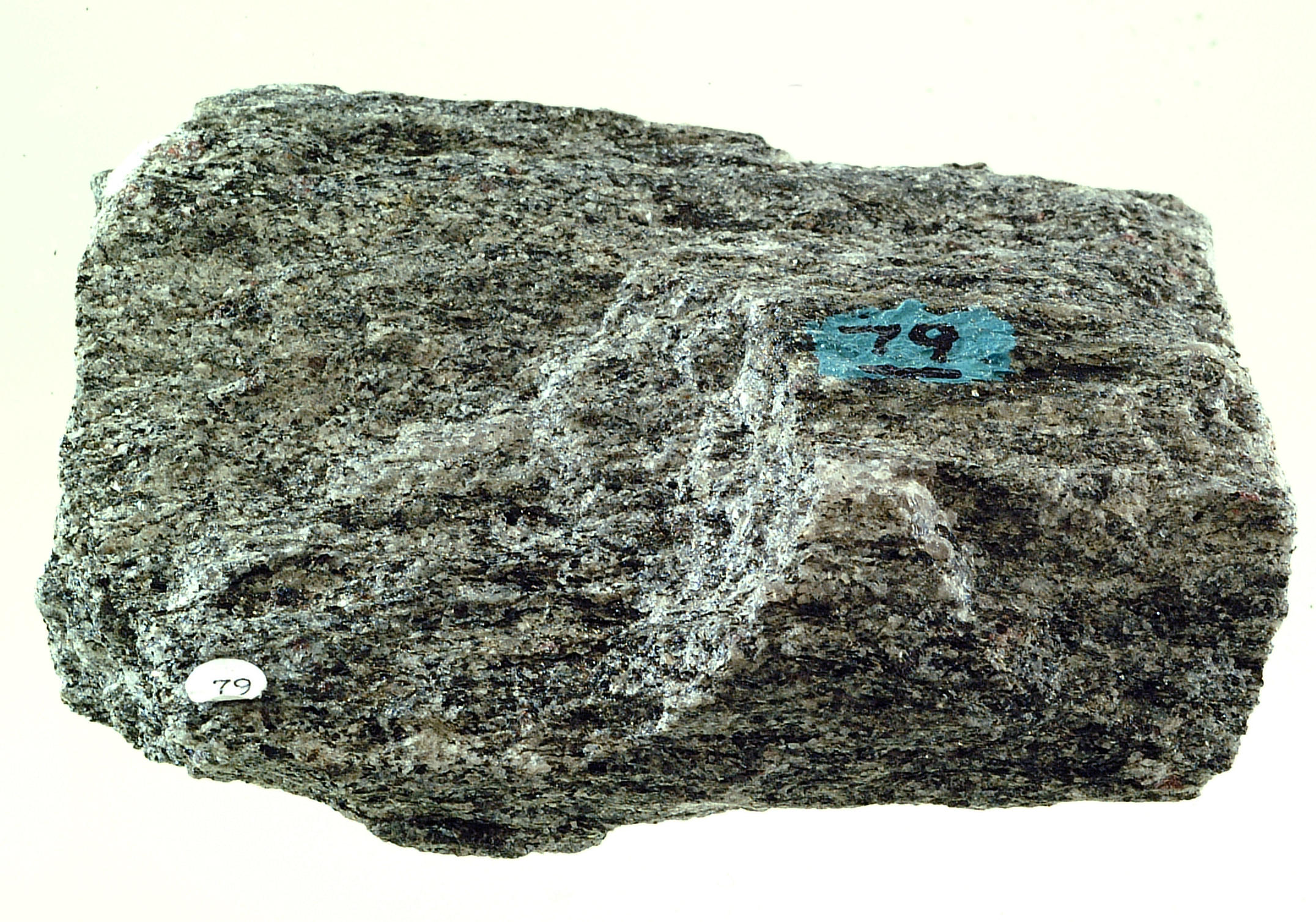
▪Phyllite
|
|
|
|
This phyllite comes from Orange County, Vermont. It contains mostly quartz, with lesser amounts of sericite, chlorite, graphite, and magnetite, but the mineral grains cannot be seen in the hand specimen. The rock has a well developed planar cleavage, and flat surfaces display a reflective phyllitic luster.
In thin section (PP view), clear quartz dominates. Long skinny stringers of muscovite and chlorite are apparent but really only identifiable by switching back and forth between PP and XP light. Minor opaque minerals are also present. In crossed polars, quartz has its normal 1st-order colors, and the skinny muscovite grains give a hint of 2nd- or 3rd-order colors. Small patches of calcite have high-order, but washed out, interference colors.
▪Phyllite
|
|
|
|
This phyllite comes from Mohave County, Arizona. It is a very fine-grained rock with a shiny phyllite luster. The rock experienced some deformation, so has a wavy/crinkly texture on its surface. The waviness is apparent in both the hand sample and the thin-section views.
The PP thin-section photo shows foliation with clear quartz layers alternating with graphite-rich layers. In crossed polars, some small skinny flakes of muscovite with 2nd- or 3rd-order colors can be identified. A few blocky patches of calcite are also present.
▪Chlorite Schist
|
|
|
|
| ⇒Link to 3D rotatable image of a chlorite schist |
The chlorite schist seen in the hand sample above comes from Jarrettsville, Maryland. It is a typical chlorite schist – green and very fine-grained. Figure 16.154 is a photo of a different chlorite schist from Michigan’s Upper Peninsula.
The thin-section views above are of a different chlorite schist. This schist contains only chlorite (pleochroic from clear to light green in the plane-polars photo) and magnetite. In crossed polars, the chlorite has low 1st-order colors.
While we commonly think of chlorite as being a low-grade mineral, sometimes that is not the case. For example, the chlorite schist seen in Figure 16.212, from Lake Martin, Alabama, contains large garnet porphyroblasts. Such porphyroblasts are common in many amphibolite facies rocks.
▪Mica Schist
|
|
|
|
This mica schist, from Gilpin, Colorado, has a wavy surface because it has been deformed. Very fine-grained muscovite gives the rock a bright sheen but individual mica crystal can only be seen with a hand lens. The hand specimen displays compositional layering when viewed parallel to the rock foliation (not seen in the hand-sample photo above). Quartz-rich layers alternate with mica-rich layers.
The thin-sections views above show the wavy deformation texture clearly. Most of the PP view is clear muscovite, but minor brown biotite is intimately mixed with the muscovite. Blocky equant clear quartz crystals are also present but are more easily distinguished from the muscovite in the XP view. In the XP view, quartz has normal 1st-order interference colors. Muscovite has interference colors that range up to 3rd-order. Biotite’s interference colors are masked by the biotite’s brown color.
▪Kyanite Schist
|
|
|
|
| ⇒Link to 3D rotatable image of this kyanite schist |
This kyanite schist comes from Manhattan, New York. Both muscovite and biotite can be seen in the hand specimen, and give the rock a sparkly appearance. The rock has a well developed foliation due both to the alignment of micas and compositional banding with quartz-rich layers alternating with more mica-rich layers.
In thin section, brown pleochroic biotite and clear muscovite laths/flakes are easily discerned. Other clear mineral grains are mostly quartz and plagioclase. The clear grains with very high relief (on the left side of the thin section in the PP view) are kyanite. Accessory opaque minerals are also present.
In the XP view, the kyanite has only 1st-order interference colors, as do the quartz and plagioclase. Muscovite has up to 3rd-order interference colors, and the deep color of the biotite partially masks its interference colors (which are also up to 3rd-order).
▪Staurolite Schist
|
|
|
|
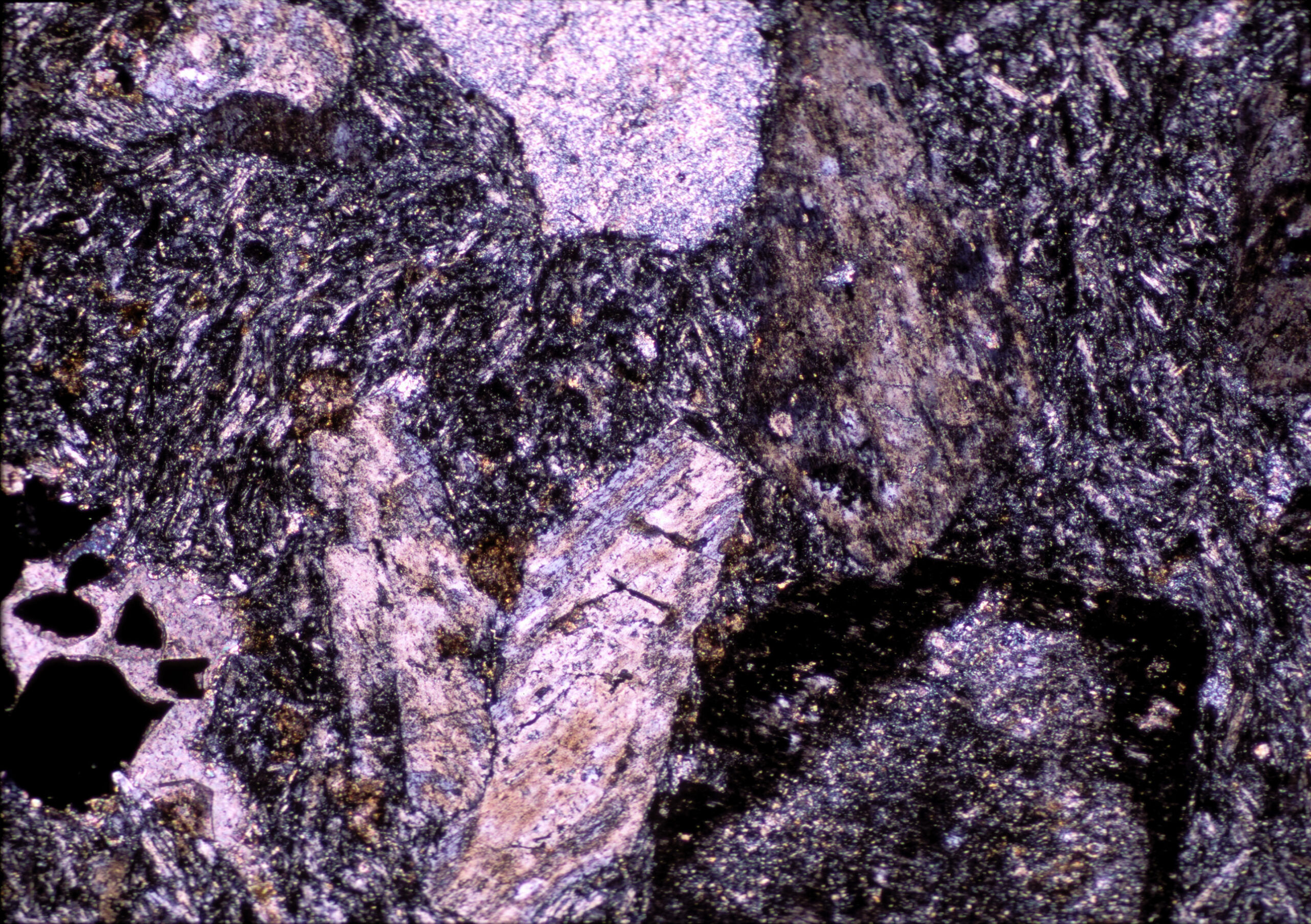
The staurolite schist seen above comes from near Gassetts, Vermont. Mica flakes up to several millimeters across give the hand specimen a sparkly appearance. Brown millimeter-sized staurolite crystals can be seen with a hand lens. Biotite and small garnets can also be seen in the hand specimen, but do not show in the thin-section views above.
In thin section (PP view), the single large staurolite crystal has its typical yellow color, good cleavage, and prismatic shape. It contains inclusions of opaque magnetite and very high-relief rutile that has a deep reddish-brown color. The clear minerals in this thin section are mostly flakes of muscovite and blocky quartz. Minor wedges of biotite are commingled with the muscovite in a few places.
In crossed polars, the staurolite has only yellow interference colors. This is typical for staurolite because the strong yellow mineral color masks any interference colors that might develop. The muscovite has up to 3rd-order colors; and quartz has normal 1st-order colors.
▪Staurolite Quartzite
|
|
|
|

This is a quartzite, from Rio Arriba County, northern New Mexico, that contains staurolite and biotite besides quartz. The rock was probably originally made of mostly light-colored quartz, but has oxidized and so now has a chocolate-brown color. This rock contains porphyroblasts of staurolite that are up to several millimeters across. The porphyroblasts are surrounded by a fine-grained quartz-mica groundmass.
In thin section, the staurolite has its typical light-yellow color and has a ragged, skeletal texture. Under crossed polars the staurolite displays yellow colors, in part because the color of the mineral masks the interference colors. The staurolite in this thin section contains lots of holes, a typical diagnostic feature for this mineral. The thin section contains abundant hematite in cracks and fractures, especially around opaque mineral grains.
The green grains (PP view) are biotite altering to chlorite. Under crossed polars, most of the biotite grains have anomalous patchy interference colors. A few small flakes of muscovite are present in the upper left corner of the thin-section views; they have 3rd-order interference colors in the XP view. Opaque magnetite is also present. Most of the thin section is quartz, and abundant hematite has filled cracks between quartz grains.
The staurolite is very hard to see in the hand samples of staurolite schist and staurolite quartzite, above. But in some rocks, it is easier to discern. The photos below show two examples. The rock on the left, from an unknown place in China, contains typical brown subhedral staurolite crystals. The rock on the right, from New Mexico, contains staurolite pseudomorphs formed from original andalusite crystals.
▪Kyanite Quartzite
|
|
|
|

| ⇒Link to 3D rotatable image of this kyanite quartzite |
The hand specimen of this quartzite, from near Ogilby, California, contains easily seen blue blades of kyanite in a sea of sparkly quartz. The quartz crystals are up to 0.3 mm across (but most are finer.) Aligned kyanite crystals, up to 2 cm long, give the rock a weak foliation.
In the thin-section view, the kyanite has high relief, good cleavage (PP view), and mostly 1st- or low 2nd-order interference colors. A few grains get to upper 2nd order (XP view). The rest of the thin section is mostly quartz. A few small grains of high-relief deep-red colored titanite are present.
In crossed polars, staurolite shows yellow, somewhat anomalous, interference colors. Quartz has it normal 1st-order colors, and abundant small flakes of muscovite (3rd-order interference colors) can be discerned.
▪Biotite Gneiss
|
|
|
|
This gneiss comes from near Cuttingsville, Vermont. Gneissic texture is well-developed. White to slightly pinkish quartz-feldspar layers alternate with gray mica-rich layers. Identifying the minerals present in hand specimen is, however, difficult because of an overall fine grain size.
In the PP view, brown biotite stands out. In places it is being replaced by green chlorite. And in some places the biotite is intimately associated with opaques that are presumably magnetite or ilmenite. The rest of the thin section is plagioclase and quartz, along with minor small opaque grains. The plagioclase appears to have a rougher surface due to incipient alteration.
In the XP view, biotite’s brown color masks its interference colors. Low-birefringence quartz is mixed with small grains of plagioclase, some of which displays albite twinning. In places the plagioclase is being replaces by sericite.
▪Garnet-Sillimanite Gneiss
|
|
|
|

| ⇒Link to 3D rotatable image of a different garnet-sillimanite gneiss |
This garnet-sillimanite gneiss comes from Washington County, New York. The rock is somewhat massive and has a quartzite-like appearance when viewed on some surfaces. It does, however, contain poorly developed gneissic banding. Red-gray layers alternate with greenish-gray layers.
In the PP thin-section view, garnet appears as blocky high-relief grains that have a very faint pinkish color. Clear sillimanite needles, many oriented parallel to the slide, have lower relief. A few squareish end views of the needles can be seen. Clear grains are mostly quartz, but minor plagioclase is present.
In the crossed-polars view, garnet is isotropic, and sillimanite has up to 3rd-order interference colors. Quartz is easily seen but plagioclase is more difficult to pick out because it does not show well-formed twins. Some of the plagioclase has altered to fine-grained high-birefringence sericite. Small grains of high-relief apatite (with 1st-order interference colors) can be seen within some of the quartz grains.
▪Garnet-Sillimanite Gneiss
|
|
|
|
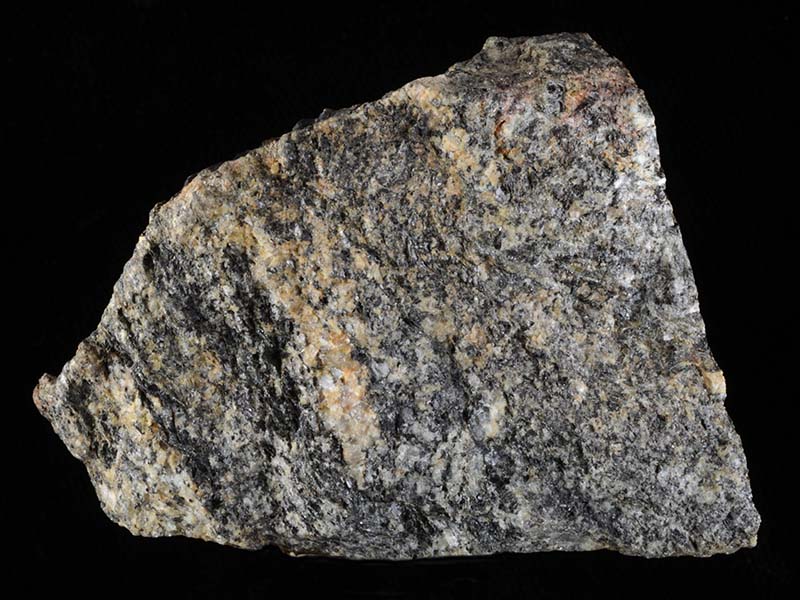

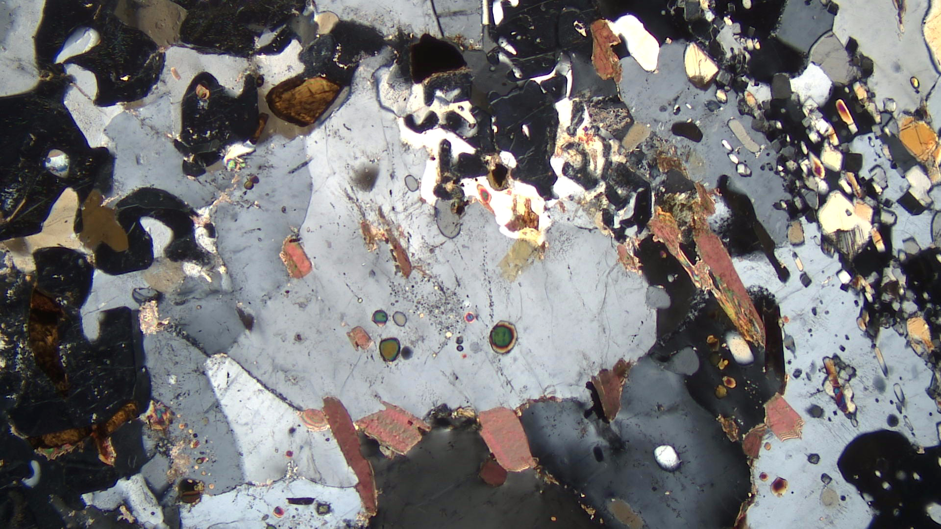
The garnet-sillimanite gneiss seen in hand specimen above comes from the Barrovian region of northeastern Scotland. The texture of the rock is part way between a schist and a gneiss. Both aligned micas and gneissic banding provide foliation. The sillimanite is not visible in this photo, but a few very small garnets can be seen associated with biotite.
The thin-section views are of a different garnet-sillimanite gneiss from near Kazabazua, Quebec. The plane-polars view shows brown pleochroic biotite, clear low-relief quartz and feldspar, and higher relief garnet and sillimanite. Several grains of deep brown rutile are present, as well as very small grains of ilmenite.
In crossed polars, quartz and plagioclase have 1st-order colors but are hard to distinguish. Garnet is isotropic, and biotite has up to 2nd- or 3rd-order colors. Sillimanite, mostly on the right side of the thin section, appears as blocky or prismatic crystals with mostly yellow-orange to 2nd-order red-purple colors. The color of rutile masks its interference colors.
▪Garnet-Cordierite Gneiss
|
|
|
|
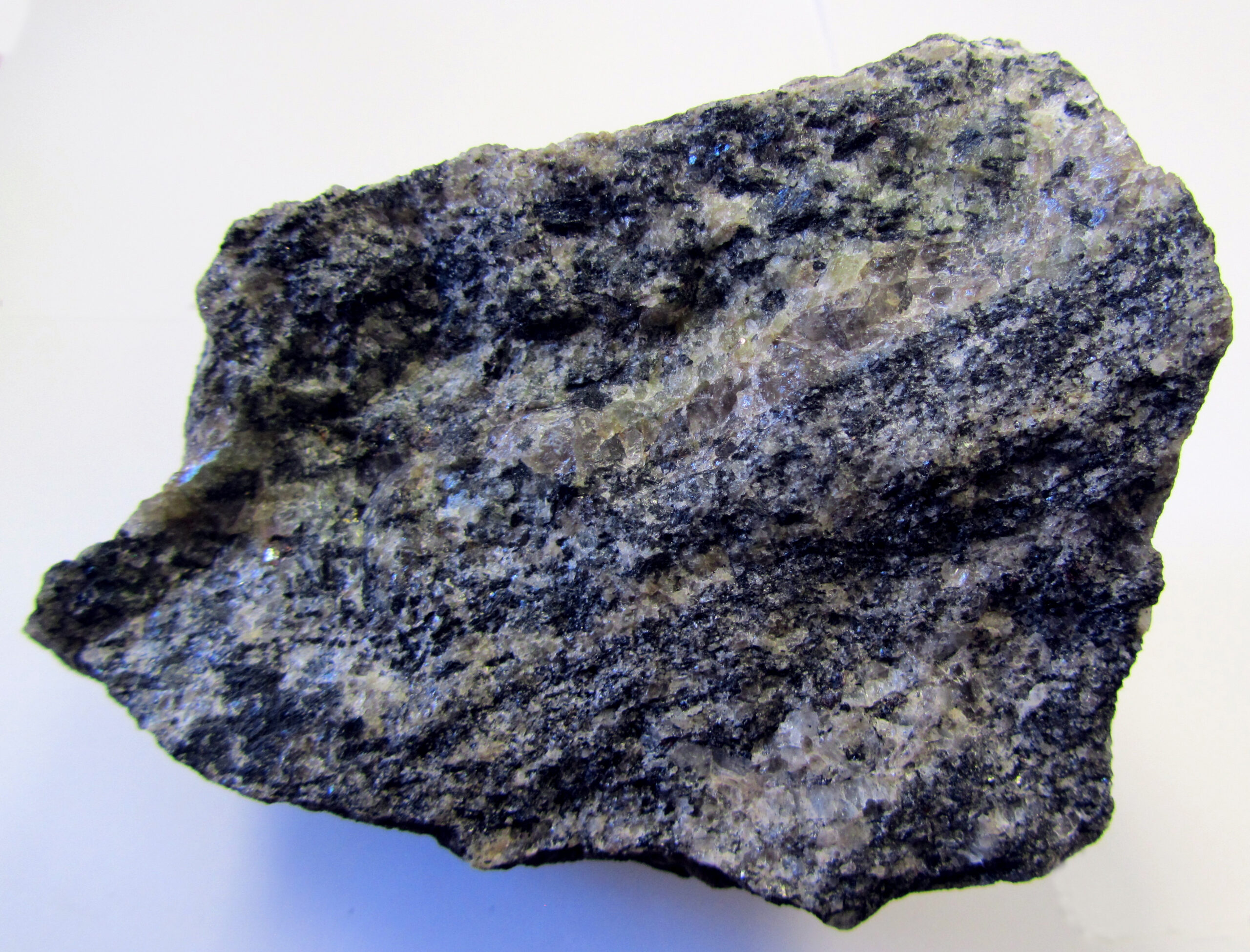
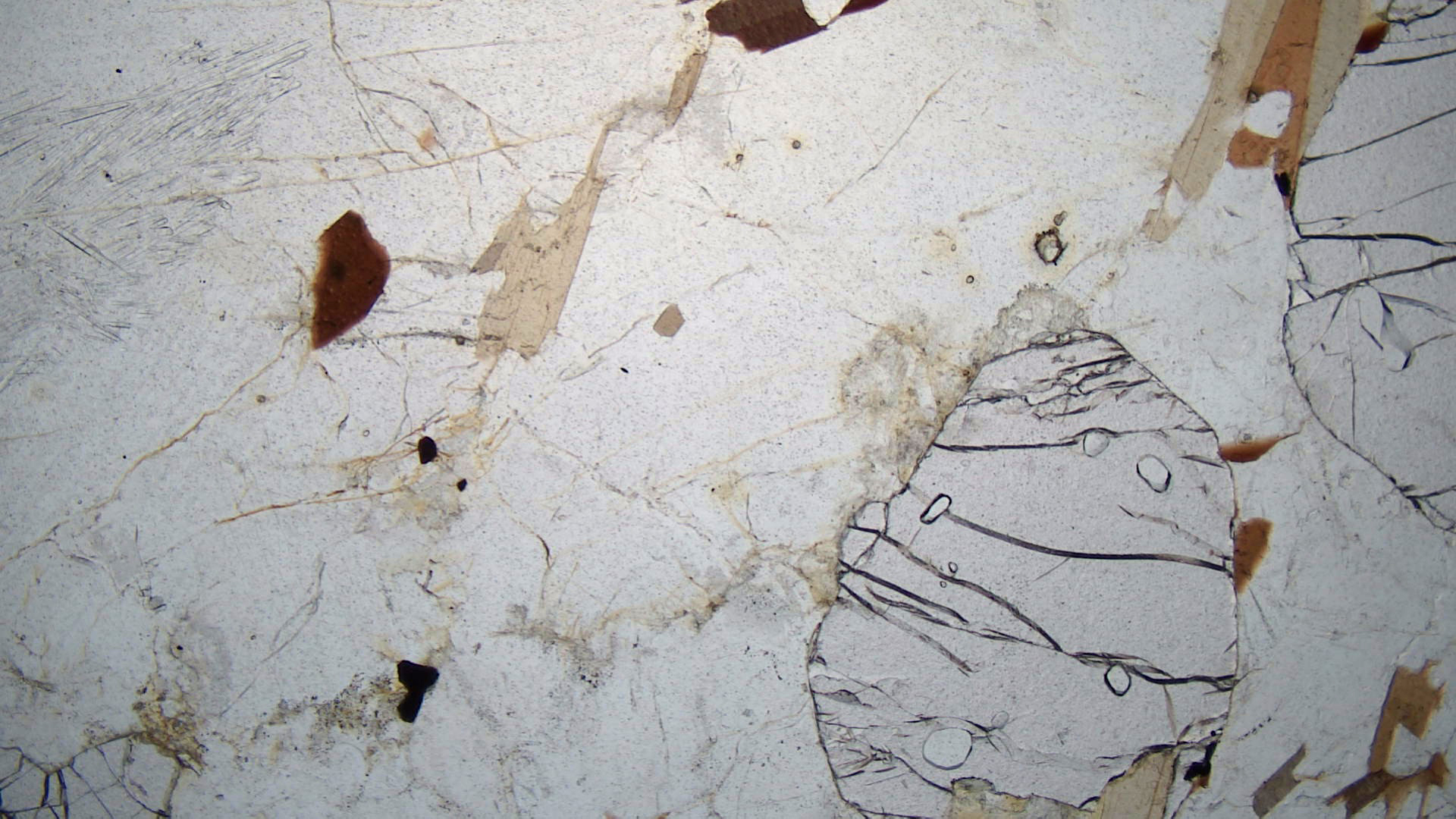
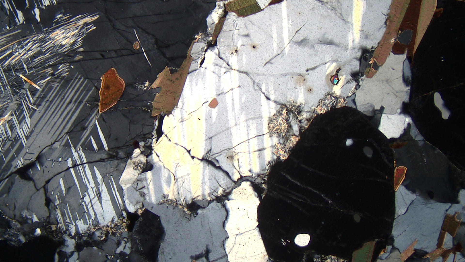
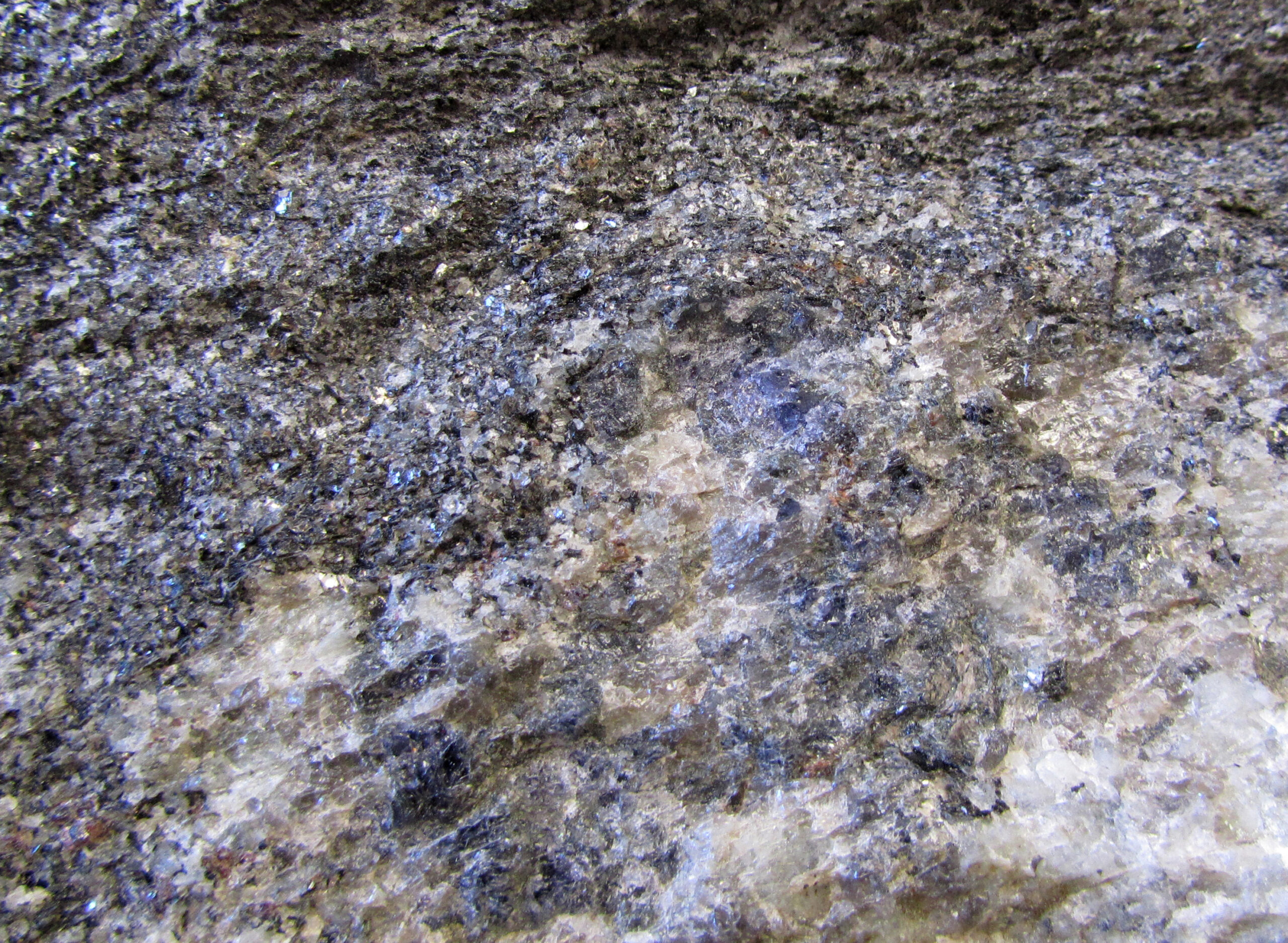
The cordierite-bearing gneiss seen in hand specimen above comes from Otter Lake, Quebec. The light-colored bands contain quartz and plagioclase, and the dark colored bands contain cordierite, biotite, and small, hard to see garnet. The rock has an overall bluish hue due to the color of cordierite, but individual cordierite grains are hard to pick out in the hand-sample photo, above. The enlarged view in Figure 16.241 shows the blue cordierite more clearly. Cordierite is quite easily distinguished when it has this blue bottle-glass appearance. Often, however, it does not and appears a drab gray color.
The thin-section views are of a different garnet-cordierite rock. In the plane-polarized view, several slightly pinkish blocky and fractured grains of garnet stand out. Some pleochroic brown mica is also present. But, most of the plane-polarized view is clear cordierite. Small sillimanite needles, enclosed by cordierite, can be seen near the upper left corner of the photo. In several places, very small crystals of radioactive zircon, mostly within cordierite, have brown burn halos around them.
In the crossed polars view, garnet is isotropic and the biotite appears pebbly green to brown. Cordierite has lamellar twins, similar to albite twinning in plagioclase. The pleochroic halos around zircon grains stand out. Untwinned clear grains are quartz. Some sericite alteration can be seen along grain boundaries.
▪Orthopyroxene-Cordierite Gneiss
|
|
|
|
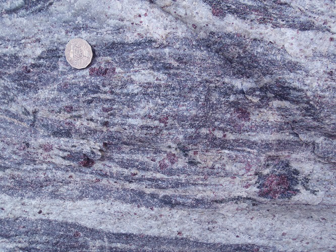

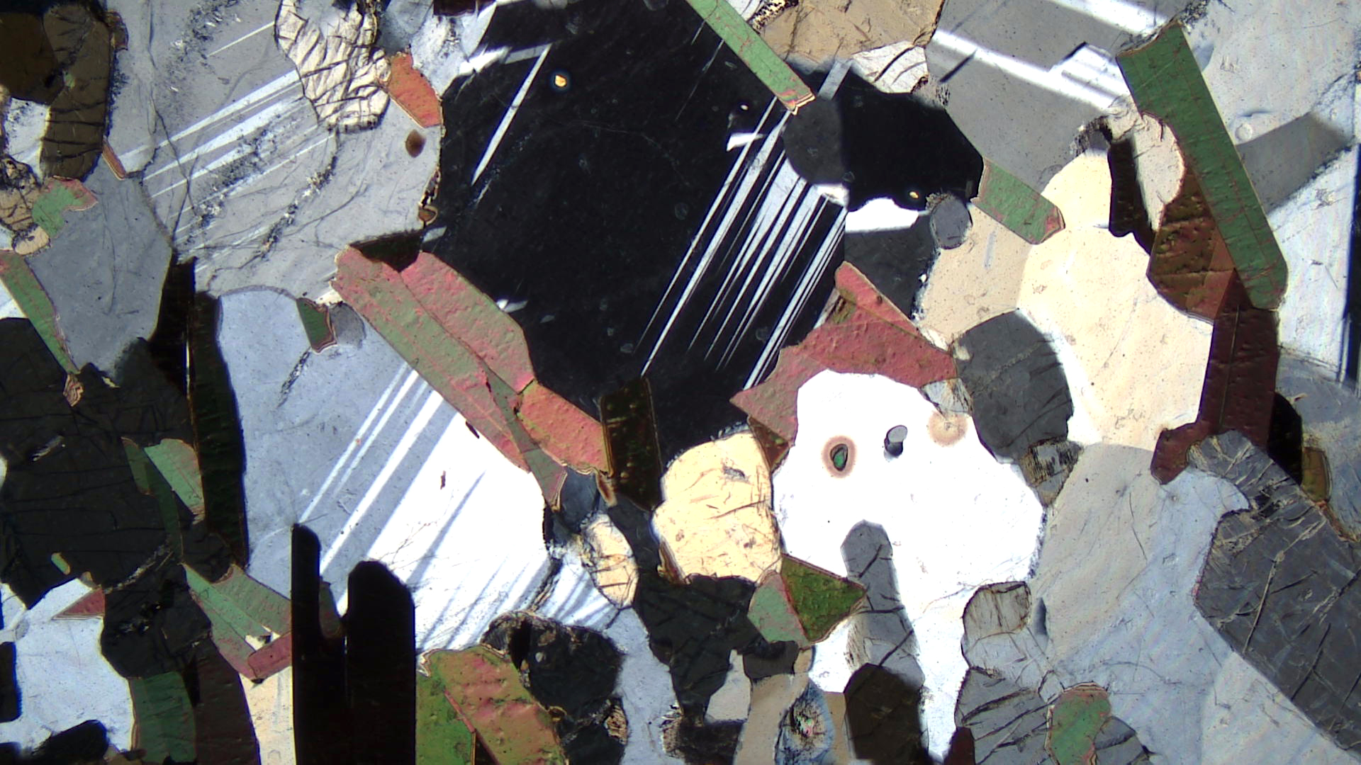
The hand sample photo above is a garnet-cordierite-orthopyroxene rock from Kottavattom, India. The thin-section views are of a cordierite-orthopyroxene gneiss from Fort Coulonge, Quebec. Other minerals present in the thin section include biotite, and zircon inclusions in cordierite. Orthopyroxene is uncommon in metapelites, and orthopyroxene and cordierite together suggest low-pressure, high-temperature metamorphism.
In the plane-polar view, orthopyroxene has high relief, shows cleavage, and is pleochroic in pink to green. Cordierite appears clear but, in places, contains conspicuous pleochroic halos (caused by radiation damage) around zircon crystals. Biotite is pleochroic in shades of brown to tan.
In crossed polars, orthopyroxene and cordierite both have 1st-order interference colors. The cordierite, however, contains conspicuous twins that look like twins in plagioclase that pinch out. Biotite has 2nd- to 3rd-order colors, and zircon even higher-order colors. Some of the pleochroic halos around zircon inclusions stand out very well.
16.3.5 Marbles and Related Rocks
In this section, we look at marbles and related rocks. This means we are considering rocks that formed from metasediments that contained significant amounts of carbonate minerals. The protoliths may have been limestones or dolostones consisting only of calcite or dolomite, or marls that contain other minerals besides carbonates. Like metabasites and metapelites, these calcareous rocks develop different minerals at different grades of metamorphism. Chapter 15 discussed all the different minerals that can form. In this chapter we look at photographs of rocks that contain the most common of those minerals. The figure below shows them listed in general order of increasing grade.
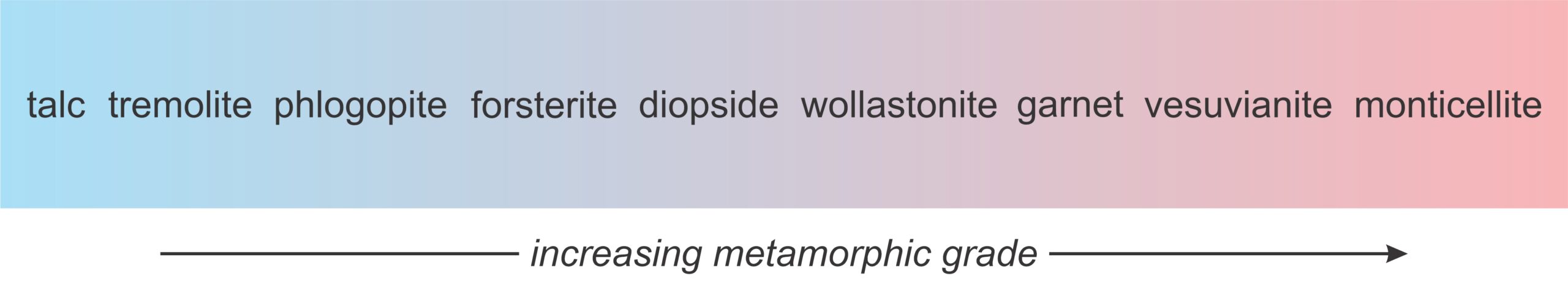
Talc, tremolite and phlogopite are hydrous minerals that give way to anhydrous diopside, wollastonite, garnet, and other minerals at high grades. But the minerals that form depend on the composition of the protolith. If the original rock was a limestone that contained no silica, none of these minerals can form. With this caveat in mind, the photos below begin with relatively low-grade marbles and end with high-grade calcsilicate rocks that contain garnet, vesuvianite, and monticellite.
▪Marble
|
|
|
|

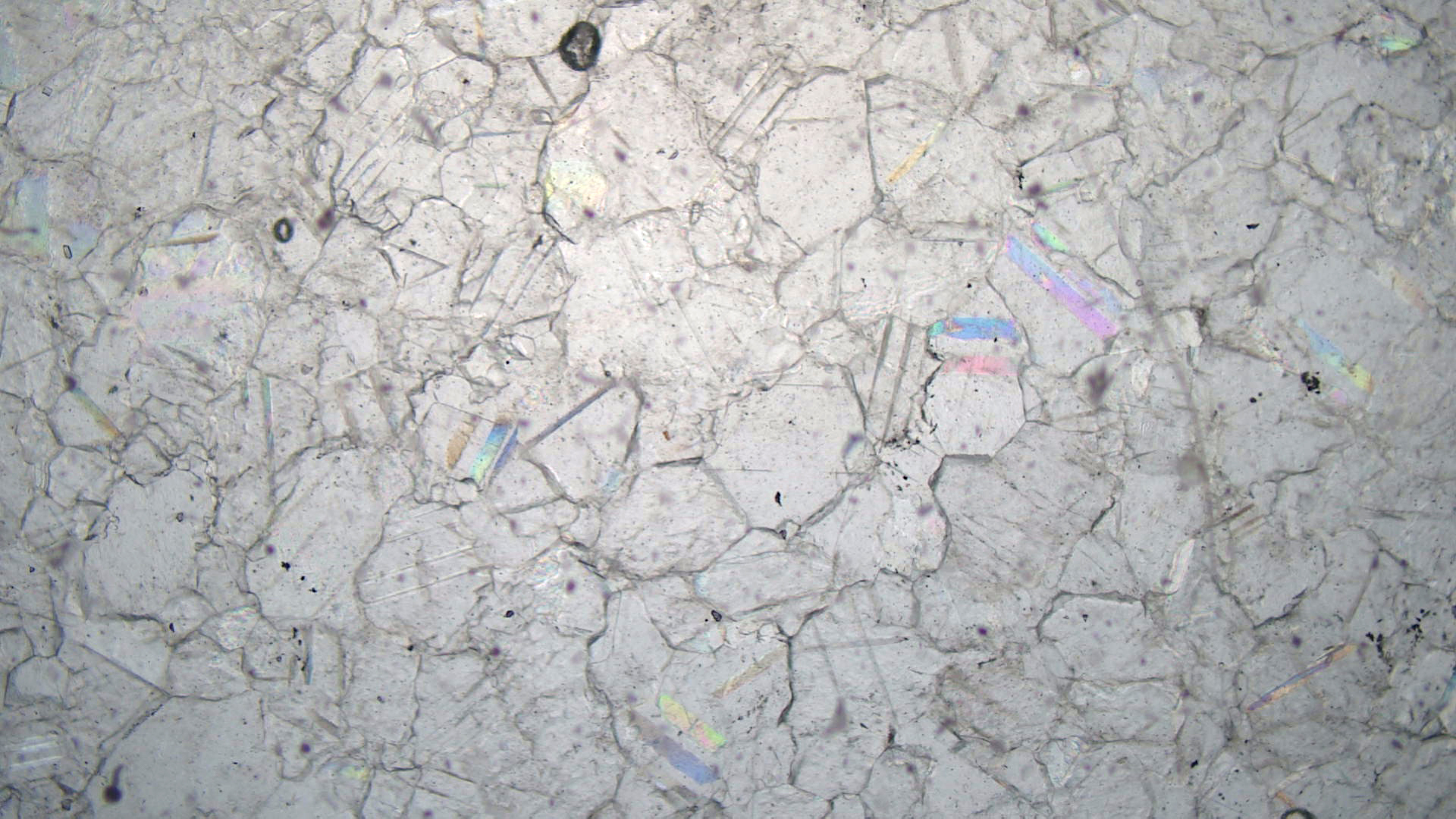
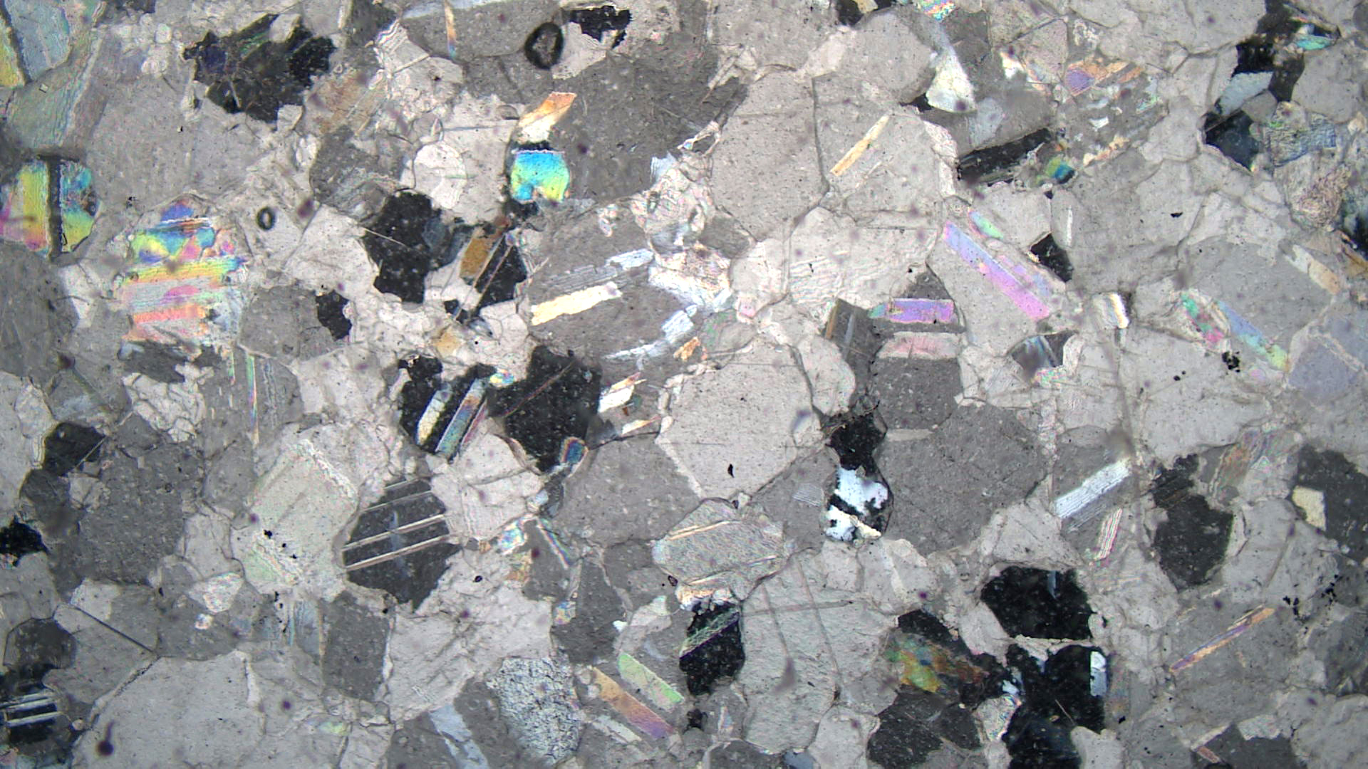
| ⇒Link to 3D rotatable image of this marble |
The marble, seen above in hand specimen and thin section, comes from Lucerne Valley, California, about 80 km northeast of Los Angeles. It contains both calcite and dolomite. The calcite is grayish with a very light blue tinge; the dolomite is lighter-colored – almost white. The two minerals are very difficult to distinguish in hand specimen and in thin section without using a chemical stain.
In the thin-section views, carbonates are the only minerals easily seen. Some of the carbonate grains in the PP view show faintly colored twins. Twins are more easily seen in the XP view. Carbonate interference colors are so high that they are very pastel or appear white in the XP view
▪Marble Containing Graphite and Phlogopite
|
|
|
|
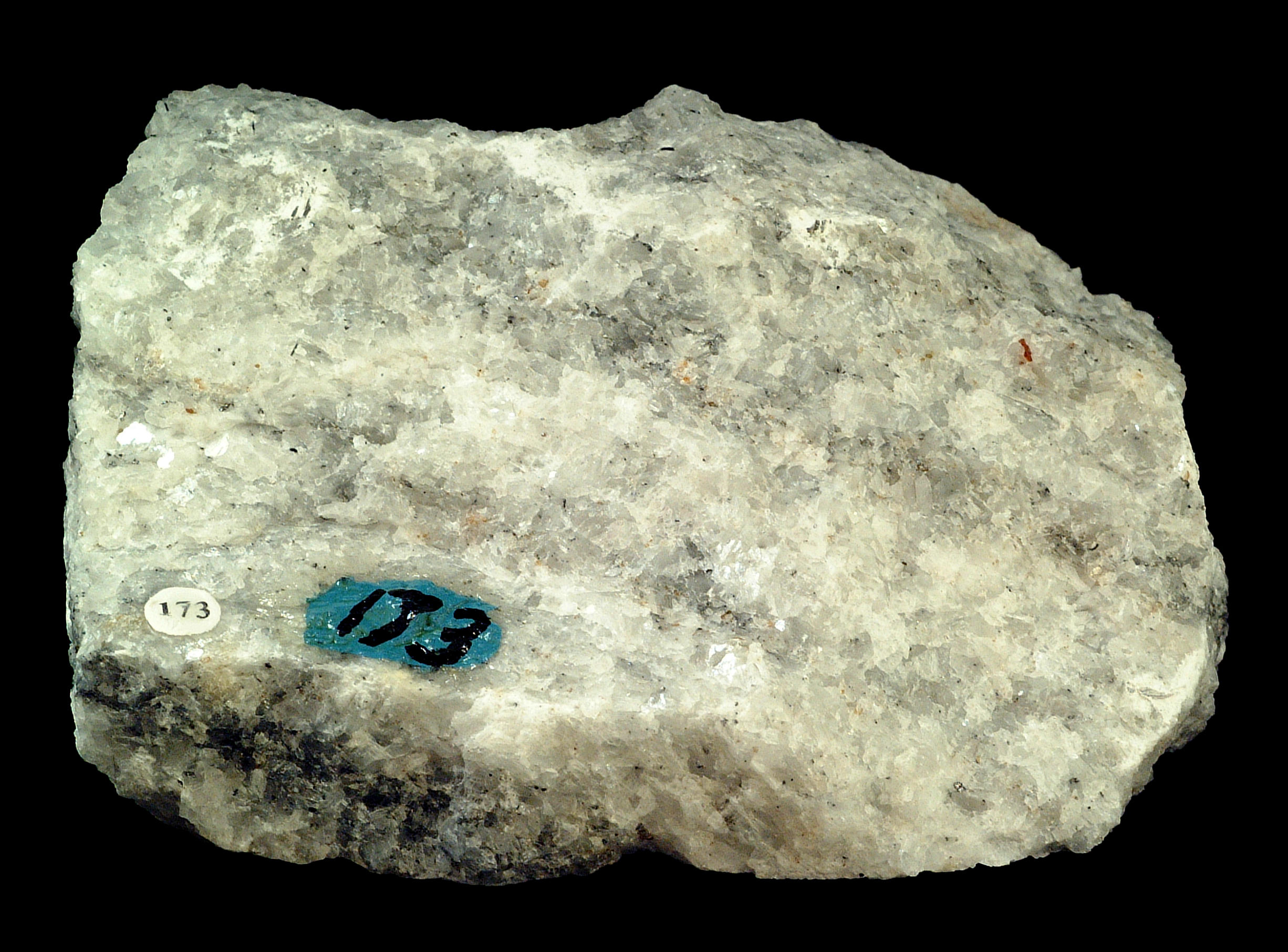
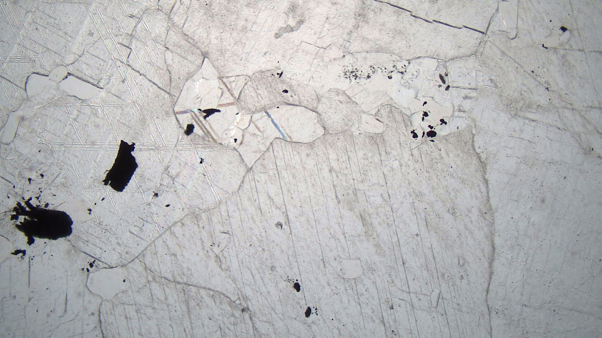

This marble comes from the western Adirondack Mountains, near Gouverneur, New York. It is mostly calcite, but close examination of the hand specimen reveals a few brown flakes of phlogopite and some silvery specks of graphite. Some marbles contain conspicuous amounts of aluminous metamorphic minerals. This one does not because it formed from metamorphism of a limestone that contained little but calcite. So the chemical components needed to form Al-rich metamorphic minerals were not present.
In the plane-polarized view, calcite is clear and shows twins. Flakes of graphite appear opaque. Some small flakes of phlogopite are present but not easily found (because they have almost no color) without switching back and forth to the crossed-polars view. In crossed polars, the calcite has its usual pastel colors. Phlogopite has up to 2nd-order blue and red interference colors. The graphite is opaque.
▪Phlogopite Marble
|
|
|
|

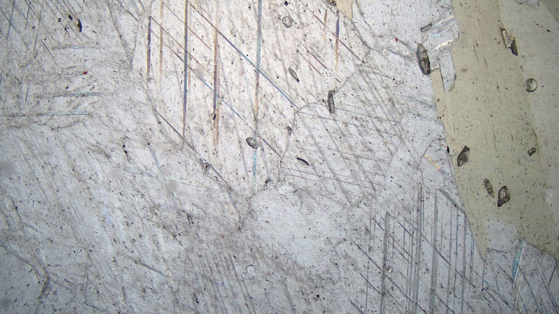
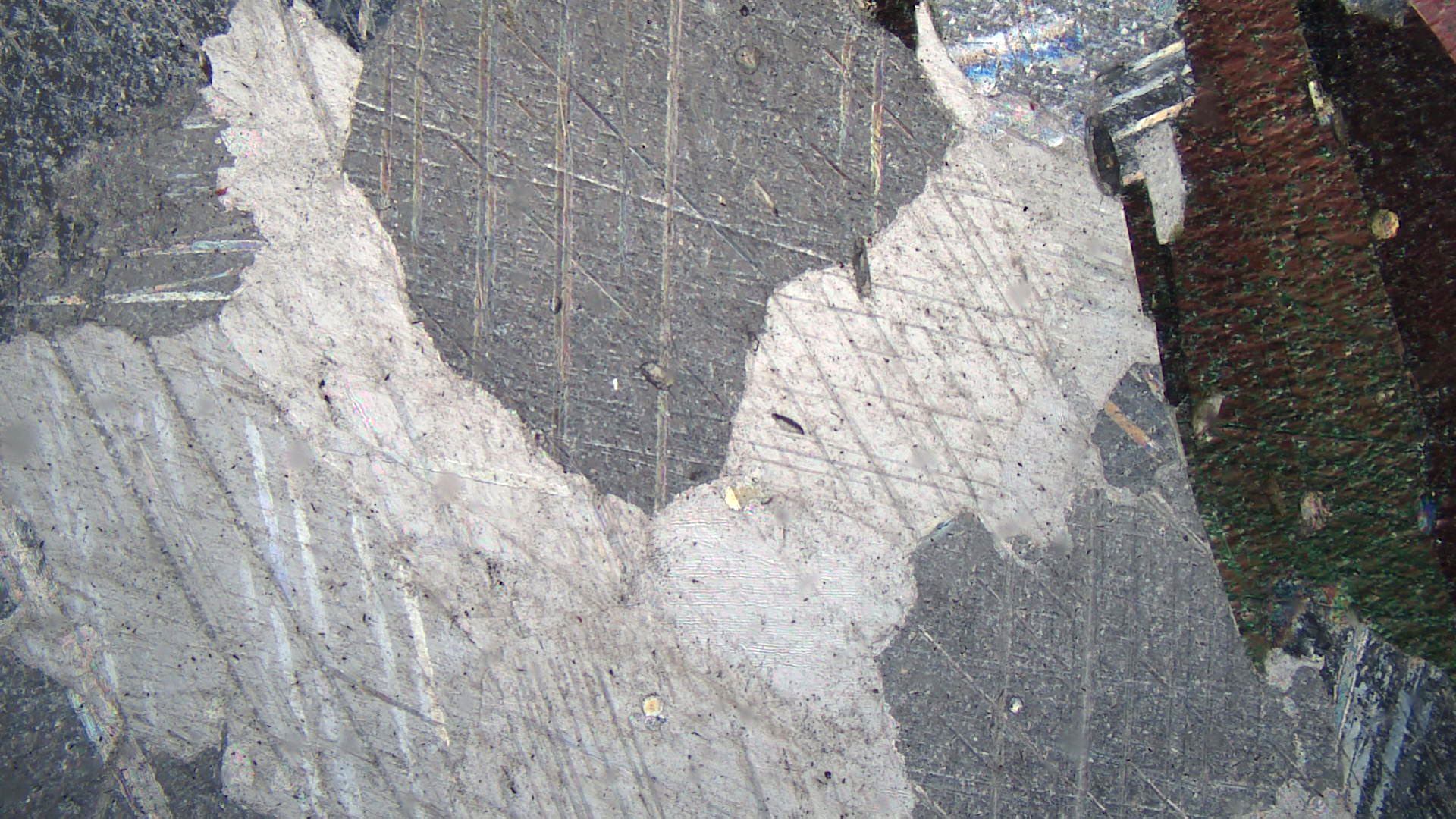
This marble, from Tate, Georgia, comprises mostly pink calcite. Darker bands in the rock contain opaque minerals and phlogopite.
In thin section, the plane-polars view reveals mostly clear calcite (some exhibiting twinning) with one large nearly vertical flake of light-greenish phlogopite on the right. A dozen or so high-relief 1-2 mm long grains of prismatic titanite are present – mostly associated with the phlogopite. A few grains of apatite (very hard to discern) and scattered very small grains of an opaque mineral can be seen.
In crossed polars, the calcite has its typical high-order pastel colors. The phlogopite exhibits a pebbly texture and 2nd-order colors.
▪Talc-Tremolite Schist
|
|
|
|
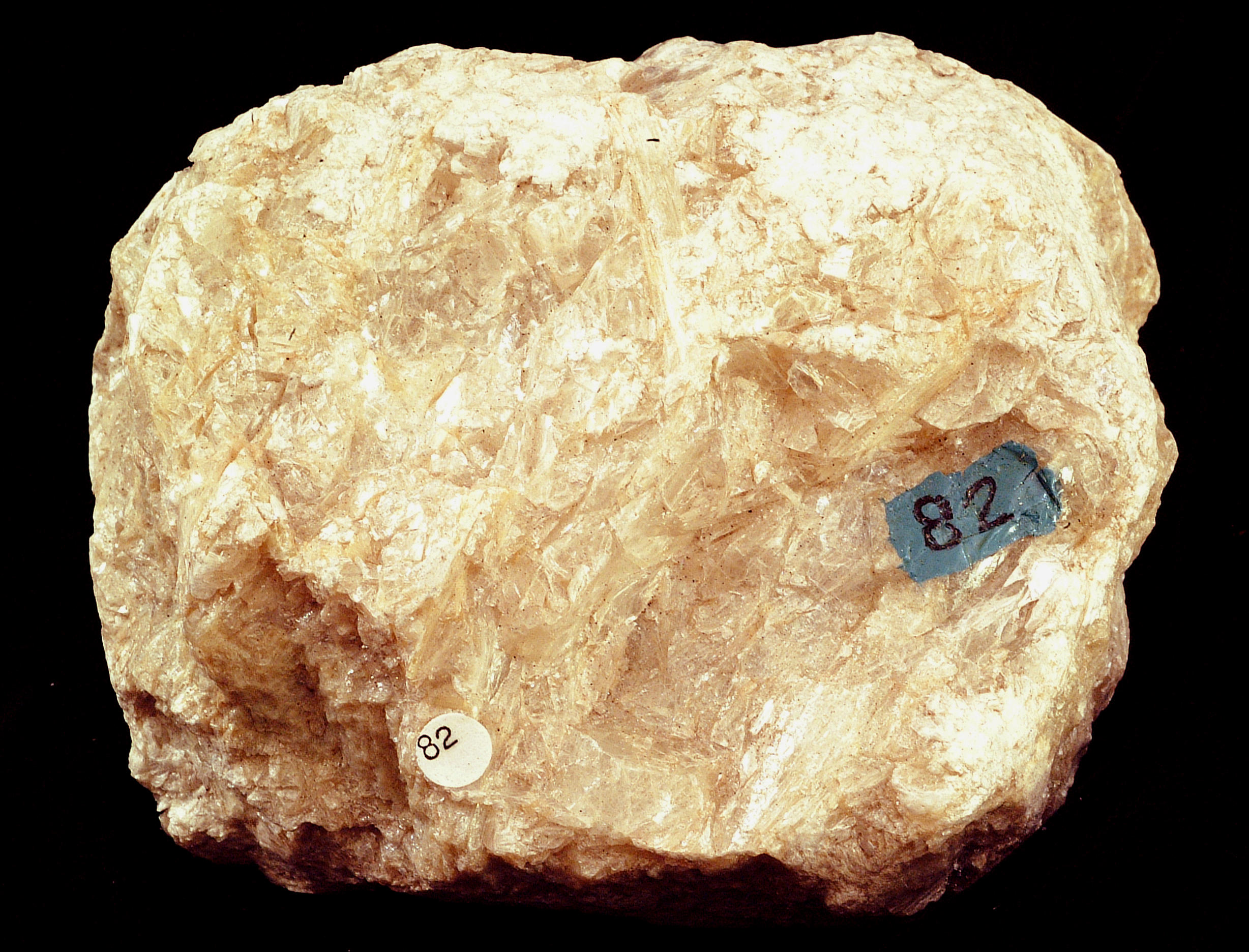

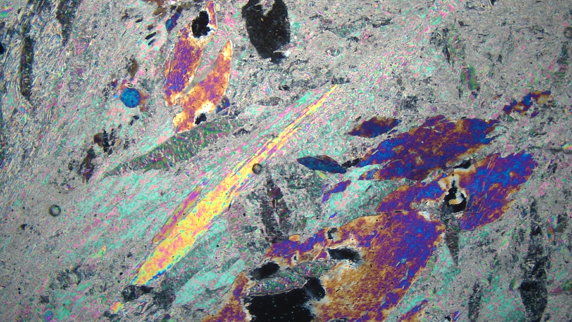
This talc-tremolite schist comes from the western Adirondack Mountains, near Gouverneur, New York. The hand sample has a very soapy feel, due to talc. A hand lens reveals sheets of talc and needles of tremolite up to 1 or 2 mm across.
In the PP thin-section view, tremolite is clear and talc has a slightly yellowish hue. One large needle and several blocky grains of tremolite are easily seen. The large tremolite grain in the lower right shows a hint of 60-120o cleavage angles (perhaps more easily seen in the XP view).
In crossed polars, tremolite blades/needles have up to 2nd-order yellow interference colors. The blocky tremolite grains have up to 2nd-order red-purple colors. Talc, in places showing a pebbly mica-like texture, displays 3rd-order colors, similar to those of muscovite.
▪Tremolite Marble
|
|
|
|
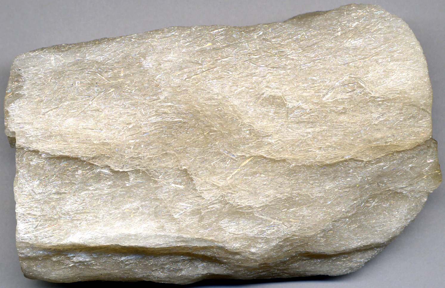
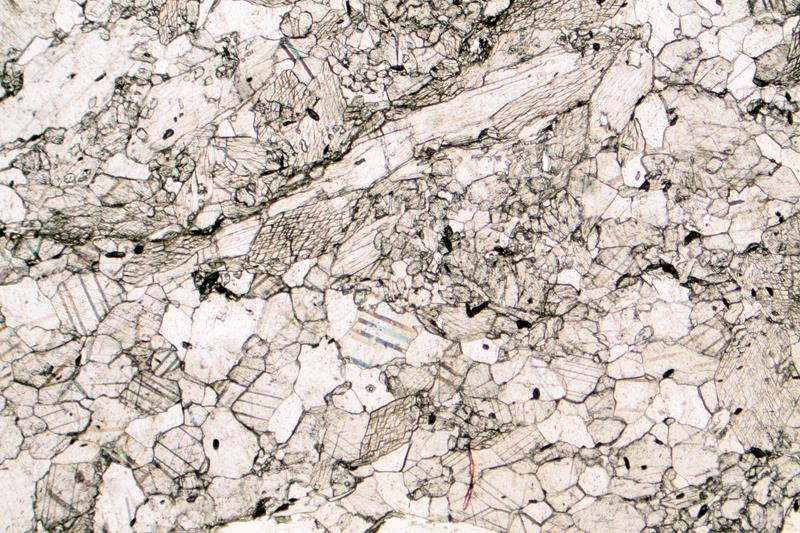
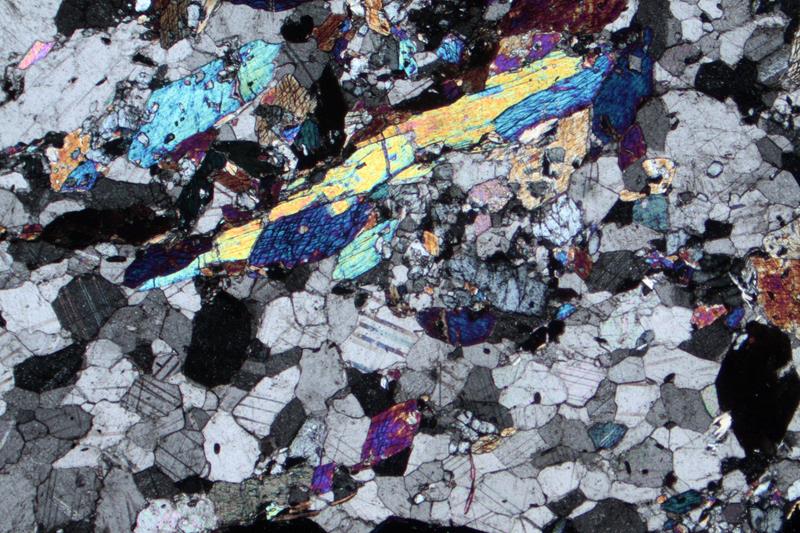
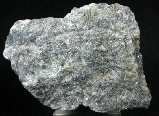
The hand sample above is a marble from the western Adirondack Mountains, New York. It contains white calcite and white tremolite. The calcite is massive and fine grained. The tremolite, a bit hard to discern, is in the form of fine needles less than 1 mm across. Some calcsilicate-rocks are nearly entirely made of tremolite; the photo seen here on the right in Figure 16.260, is an example. It contains only radiating aggregates of tremolite crystals.
The thin-section views above are of a different marble – not the same one seen in hand sample. In the plane-polars view, calcite has high relief, and twinning is apparent in most grains. Tremolite crystals are lower relief, in the form of blades or needles. End views of tremolite crystals have a diamond shape and display 60-120o cleavage angles. A few specks of an opaque mineral can be seen.
In crossed polars, tremolite blades/needles have up to 2nd-order blue and yellow interference colors; end views display 3rd-order colors. Calcite displays its typical high-order pastel interference colors.
▪Forsterite Marble
|
|
|
|
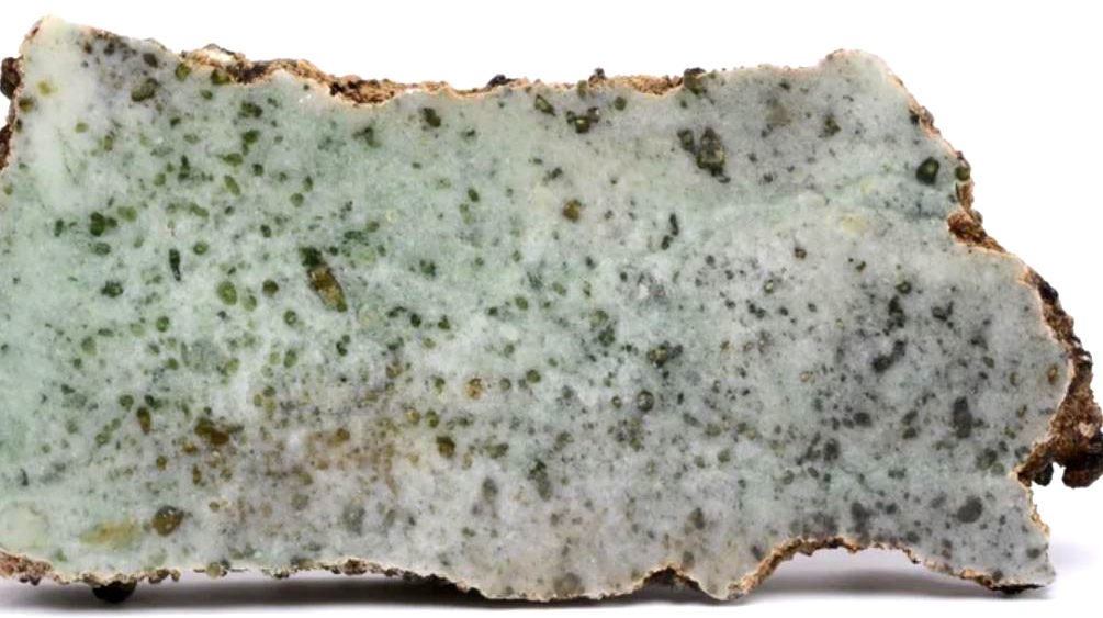
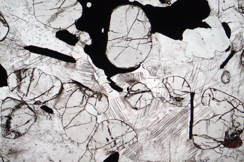
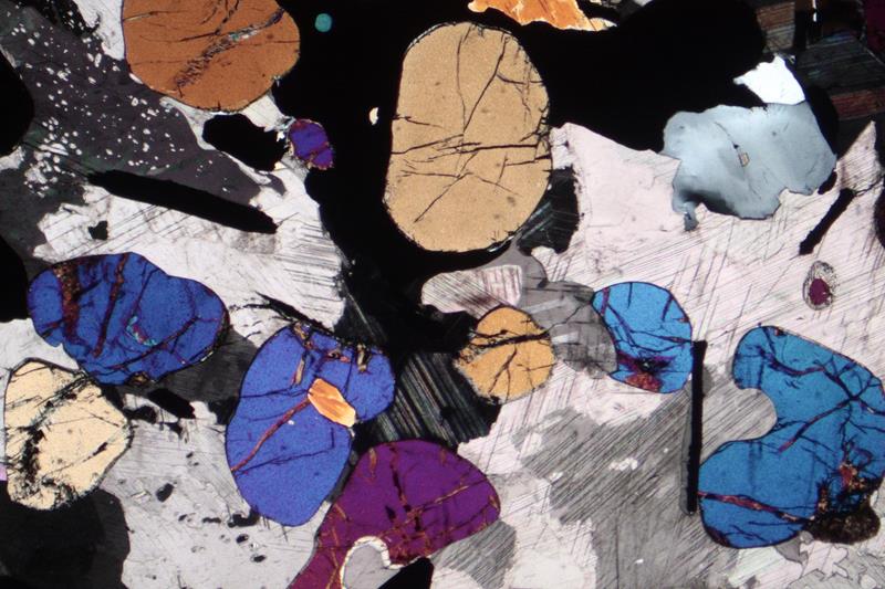
The hand sample seen in the photo above comes from near Loch Duich in western Scotland. The forsterite appears as pale green crystals that, when present, are sometimes hard to distinguish from diopside in marbles.
The thin-section views are of a different forsterite marble (from Parone, Italy). The forsterite appears as high-relief rounded grains, some quite fractured, in the plane-polarized view. The clear grains around the forsterite are all calcite, some showing a hint of twinning in the PP view. A significant amount of unidentified opaque mineral grains are also present.
In crossed polars, the forsterite has up to high 2nd-order interference colors. The twinned calcite displays high-order but hard to see pastel hues.
▪Forsterite-Diopside Marble
|
|
|
|
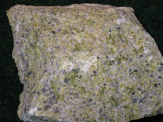

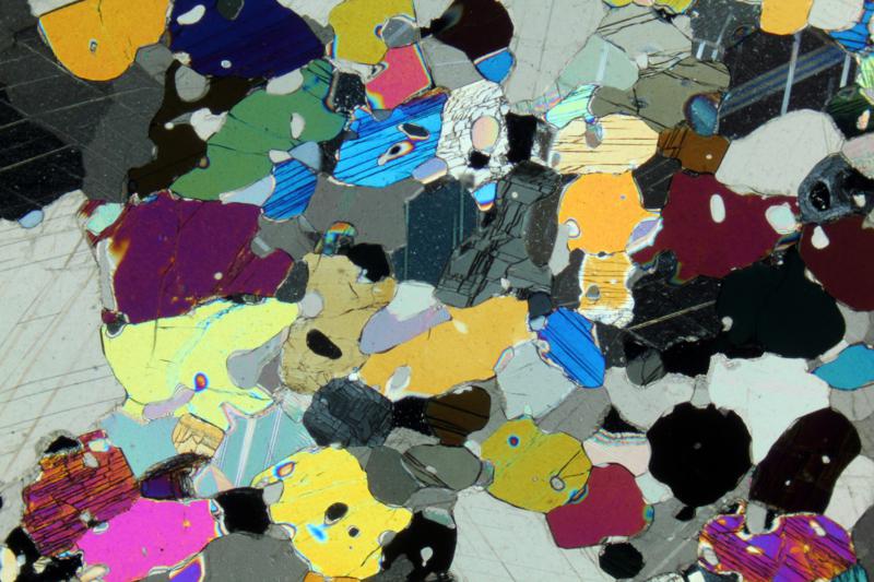
The hand sample seen above is the same as the one in Figure 15.3. It is a forsterite marble from Baffin Island, Canada. Besides light-green forsterite, it also contains lesser amounts of dark-green diopside.
In the plane-polarized view, the pyroxene grains are nearly clear, perhaps slightly greenish, and display well-developed cleavage. One millimeter-sized grain just below and left of the center of the view shows pyroxenes near-90o cleavage angle well. Calcite and olivine grains are nearly clear too, but calcite displays easily seen twinning in two directions. High-relief olivine grains are well-rounded, lack cleavage and stand out above the calcite. Some of the olivine has a faint gray color compared with the other minerals. The tremolite-rock seen above in Figure 16.260 is another example of a calcsilicate rock devoid of carbonate.
In crossed polars both the diopside and olivine have 2nd-order interference colors. The diopside has slightly greater birefringence and colors get to upper 2nd-order. Because of its very high birefringence, calcite’s interference colors cannot be easily seen.
▪Diopside Marble
|
|
|
|
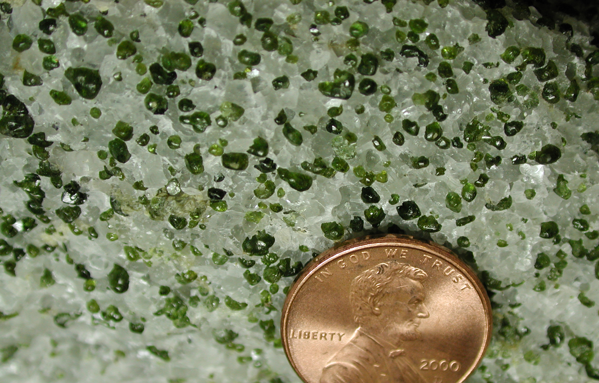
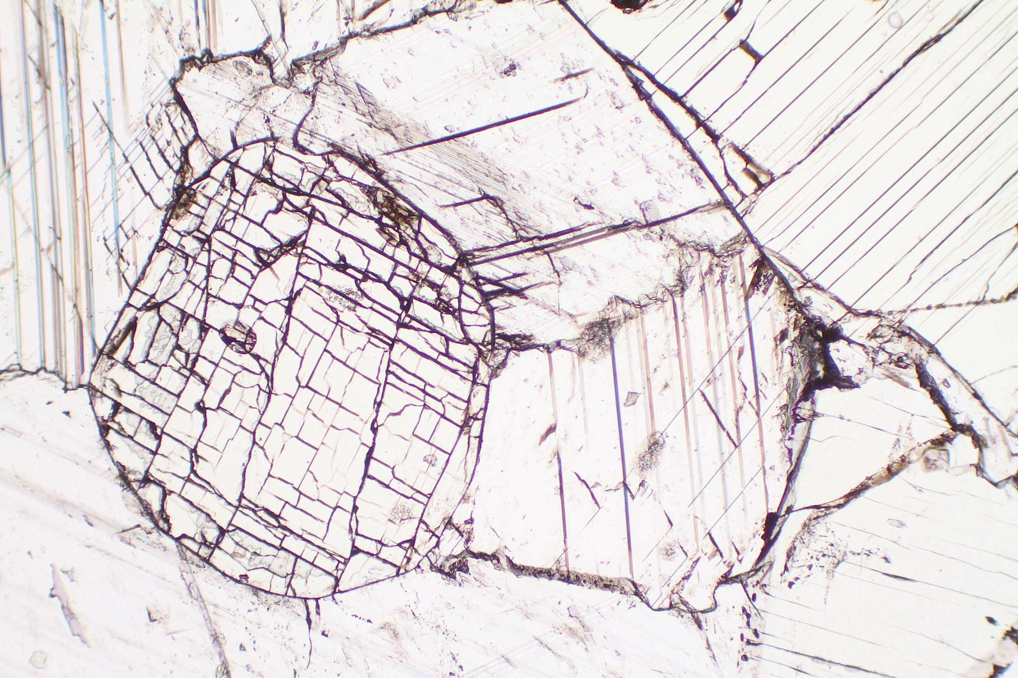
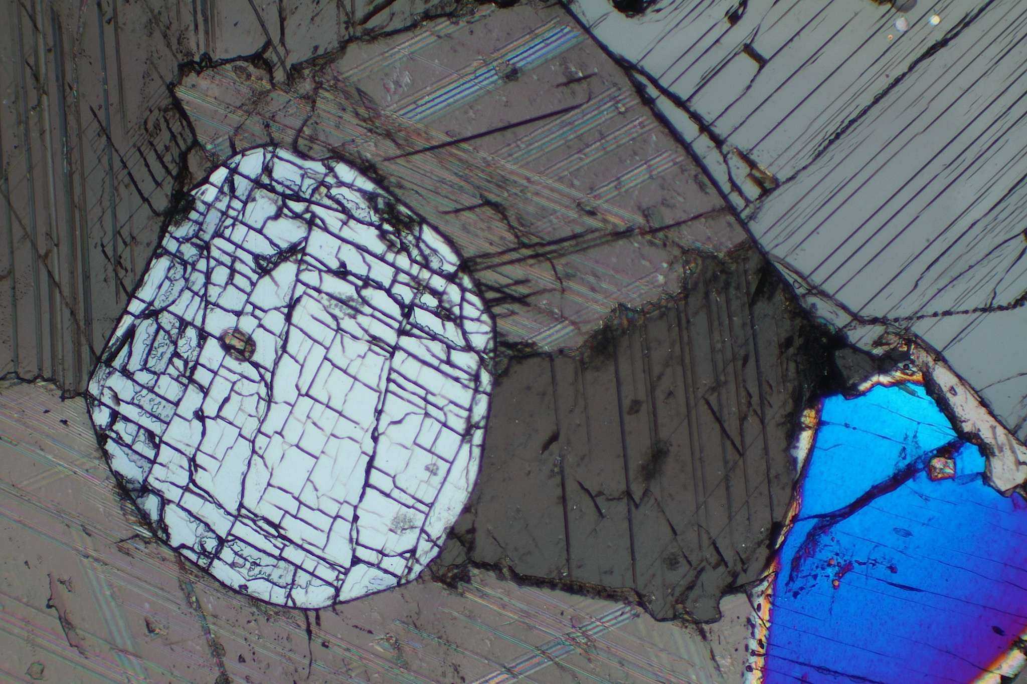
The photo on the left above is a diopside-bearing marble from Cascade Slide, in New York’s Adirondack Mountains. This marble is in a block of calcsilicate rock, partially covered, that is included within anorthosite. The exposed portion of the calcsilicate rock is about 150 m long and 20 m wide (Baillieul, 1976). This is a typical diopside marble containing green diopside crystals up to 2 mm across surrounded by white calcite.
The thin-section views above show diopside (high relief, near 90o cleavage angles) and calcite (with twins visible in both the PP and XP views). In the XP view, the diopside crystal that shows two cleavages displays minimum interference colors (gray). The other diopside crystal, that is viewed perpendicular to its optic axes, displays maximum (2nd-order blue) interference colors. Calcite’s interference colors are pale pastels of high order.
|
|
|
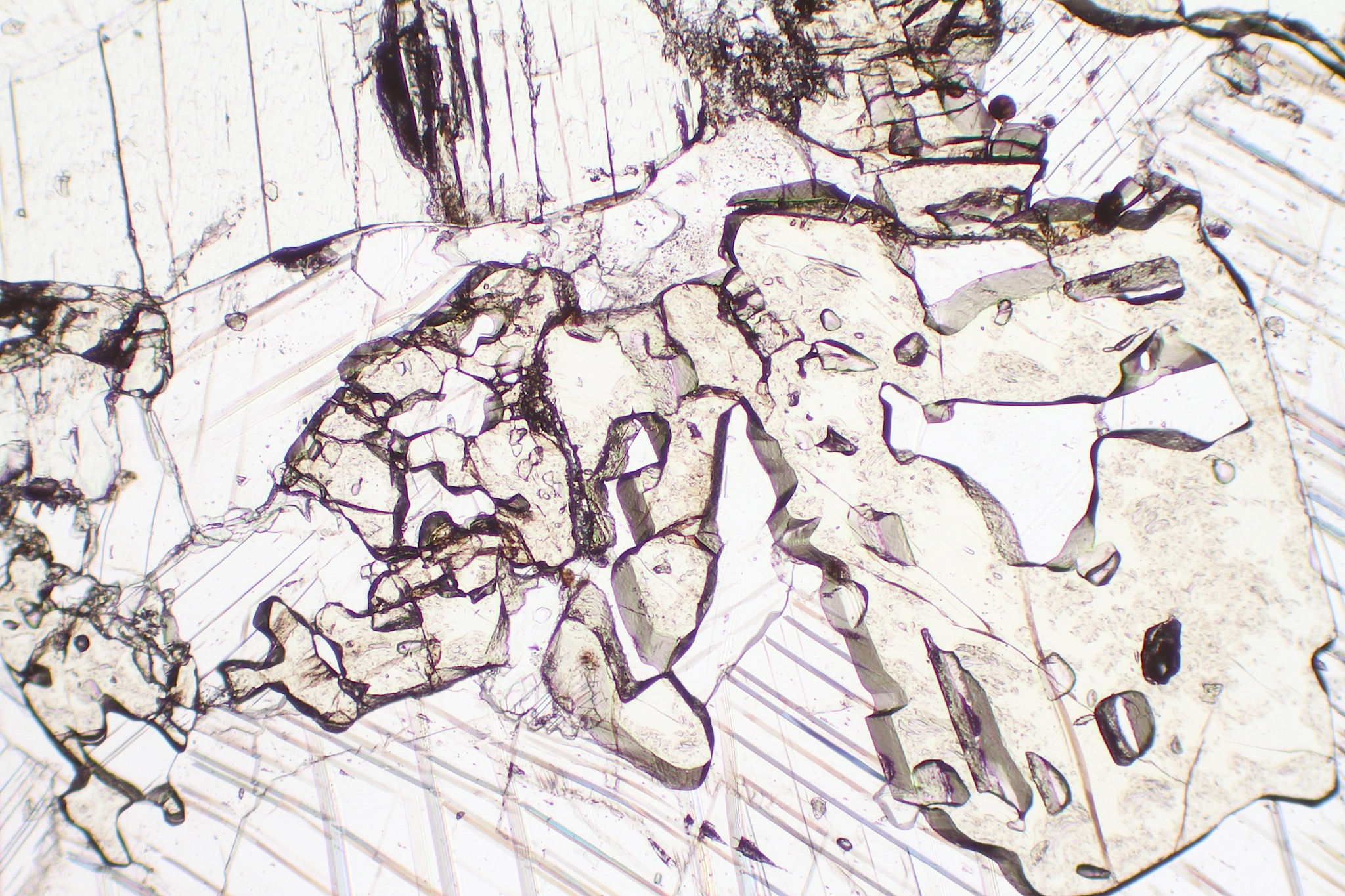
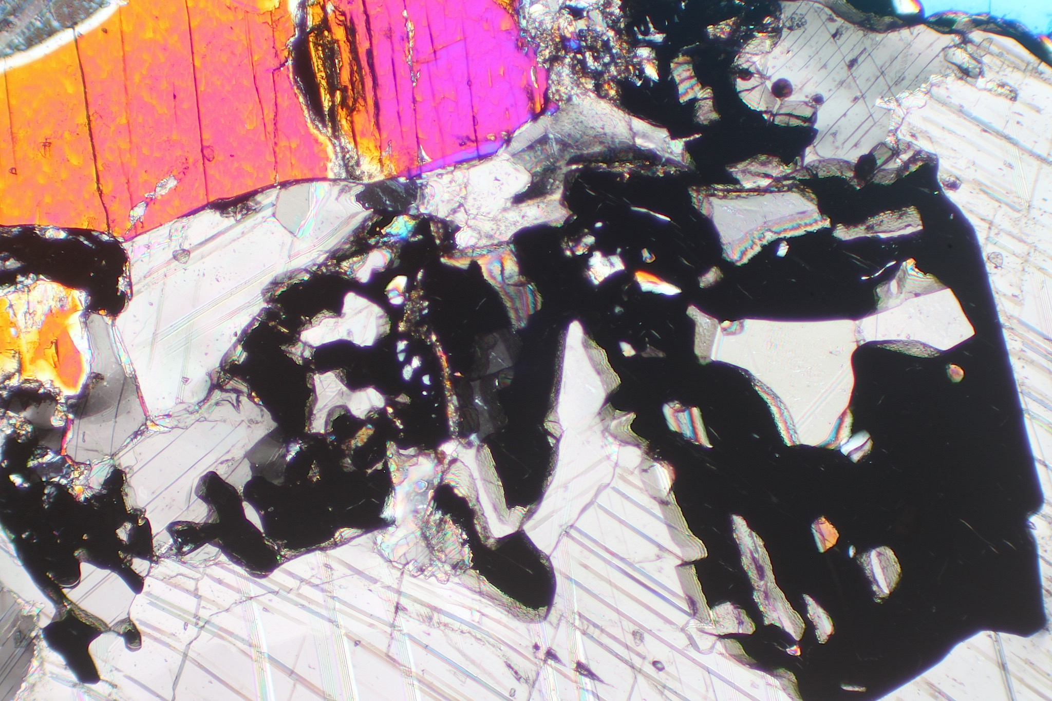
The two thin-section views in Figures 16.270 and 16.271 are of a diopside-bearing marble that contains garnet. This sample, like the previous one, comes from Cascade Slide in the Adirondacks.
The large grain in the upper left of the view, that has near vertical cleavage, is diopside. In the PP view, the diopside is almost clear. The garnet is clear, in blobby/patchy crystals, has no cleavage, but has considerably more relief than the diopside. The low-relief calcite, in the background, is clear but shows twinning. In crossed polars, the diopside displays vivid high 2nd-order interference colors. Calcite’s interference colors are so high-order and washed out that they cannot be easily seen. The garnet is isotropic.
|
|
|
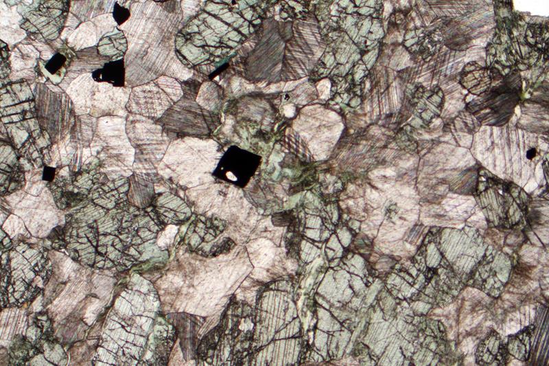
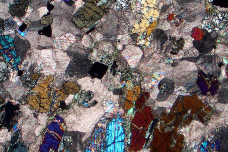
The two thin-section views on the right show a different diopside-bearing marble. This one is from Candoglia, Italy. In the PP view, the diopside has a more pronounced green color than the two diopside rocks described above. The calcite is clear to gray and displays twinning. Several opaque grains are also present. In crossed polars, the diopside displays up to high 2nd-order interference colors. Calcite’s interference colors, as usual, cannot be easily discerned.
▪Garnet-Wollastonite Calcsilicate
Calcsilicate rocks form by contact metamorphism and metasomatism of carbonate-rich protoliths. During this kind of metamorphism, coarsely-crystalline silicates typically replace some or all original carbonate minerals as metasomatism occurs. Unlike the diopside marbles we saw above, the rock seen below contains no calcite, although it surely did prior to metamorphism.
|
|
|
|
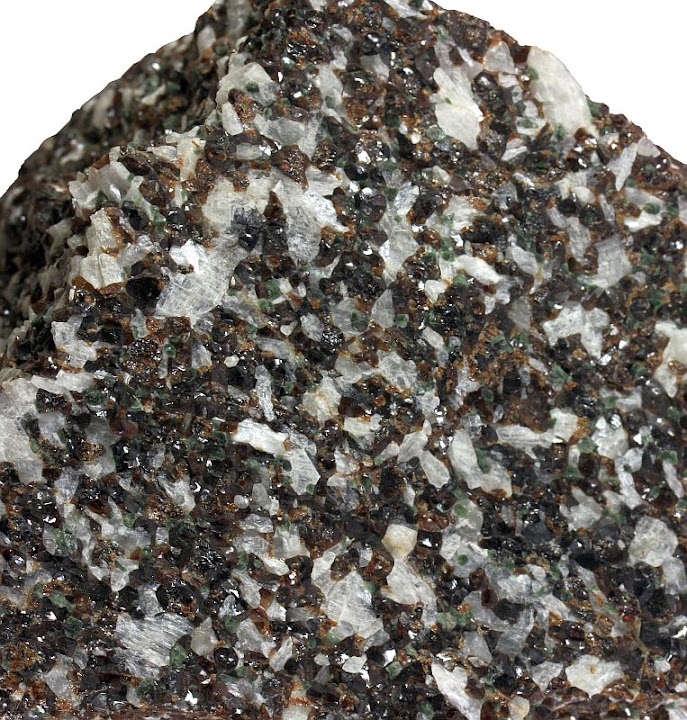

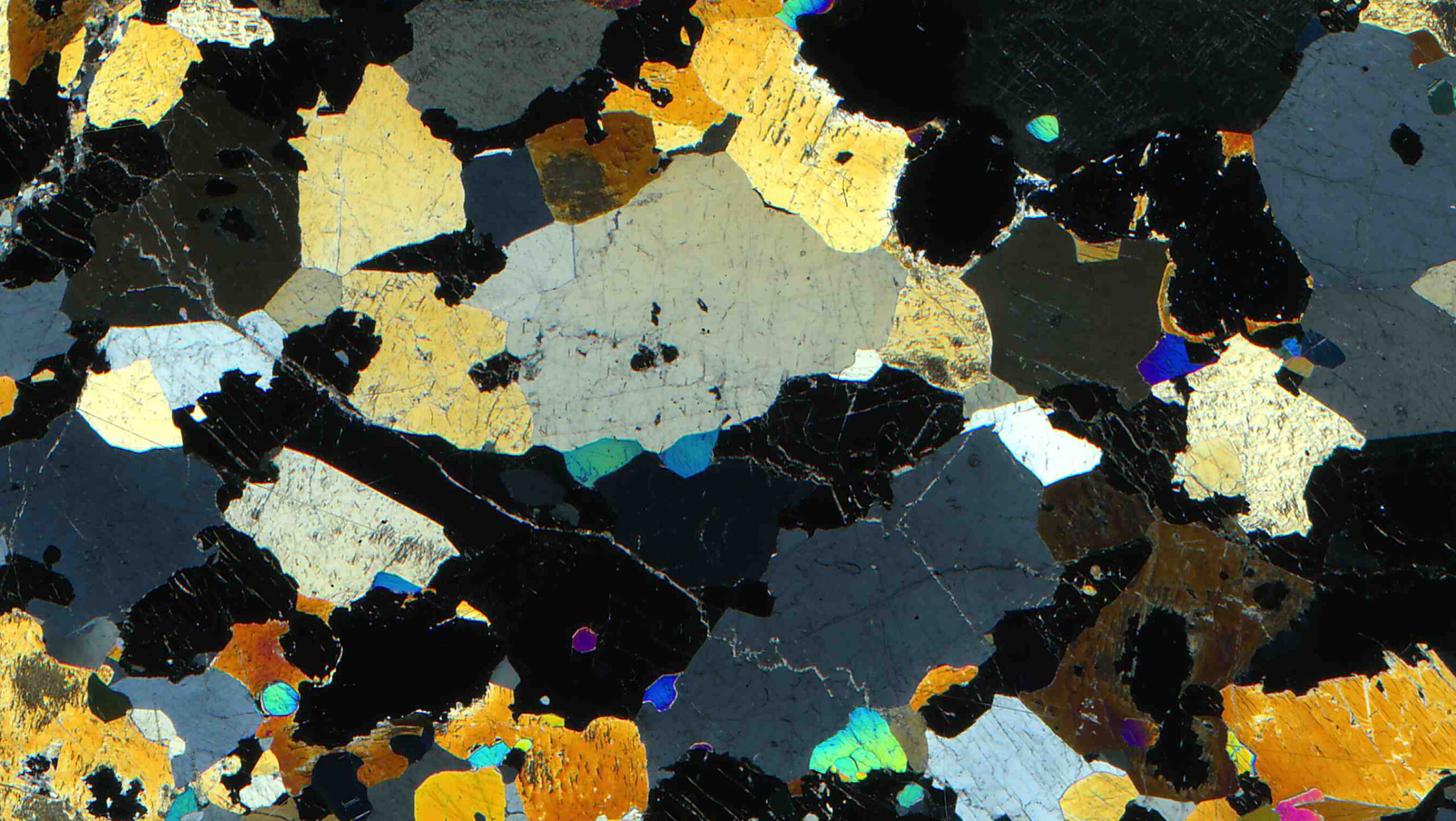
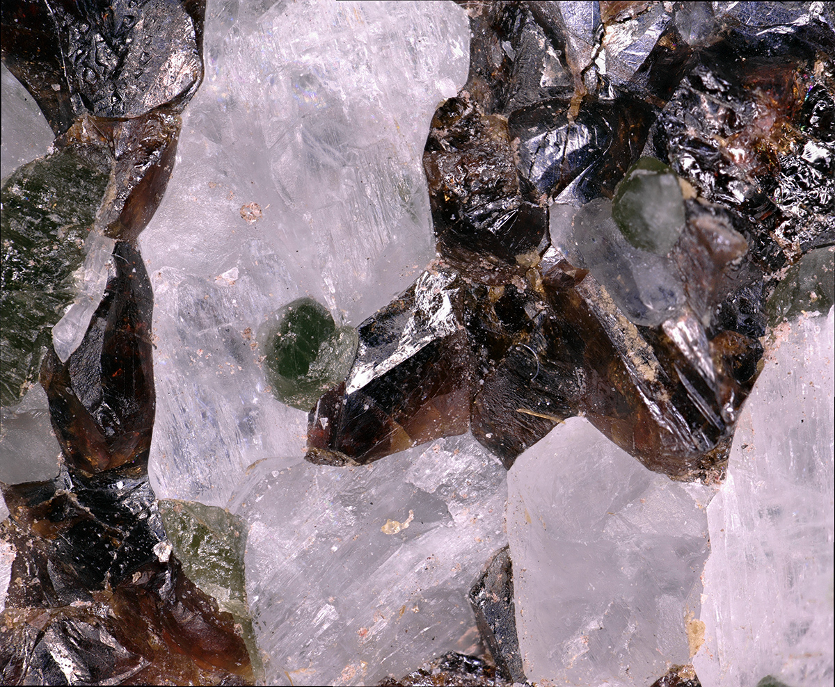
The photo in Figure 16.274 shows a garnet-diopside-wollastonite calcsilicate from the Lewis Mine, a wollastonite mine near Willsboro, New York. Figure 16.277 is a closeup view of the same rock. This classic mineral collecting site provides high-quality samples of calcsilicate rocks containing red andradite garnet, green diopside, and bladed white wollastonite.
The plane-polar and crossed-polar views above are scans that covered most of an entire thin section of a different rock from the Lewis Mine. In the PP view, the garnet has relatively high relief and a light pink color. The diopside is light green. All the clear mineral grains are wollastonite. In the XP view, garnet is isotropic, wollastonite has only up to middle 1st-order interference colors, and the diopside has up to high 2nd-order colors. Note that the two scans contain several (isotropic) holes where mineral grains were plucked during thin-section polishing.
▪Vesuvianite Marble
|
|
|
|
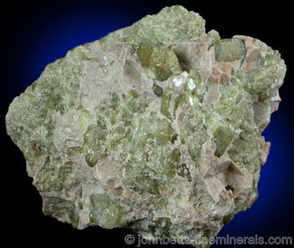
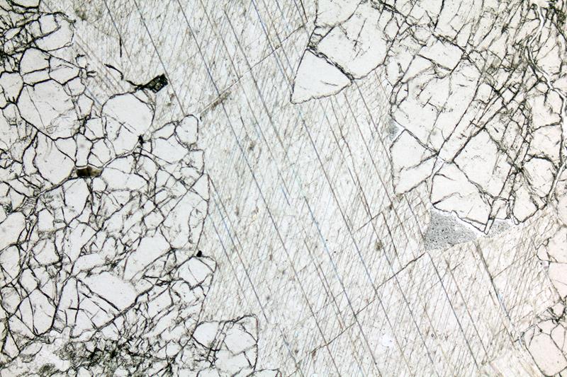
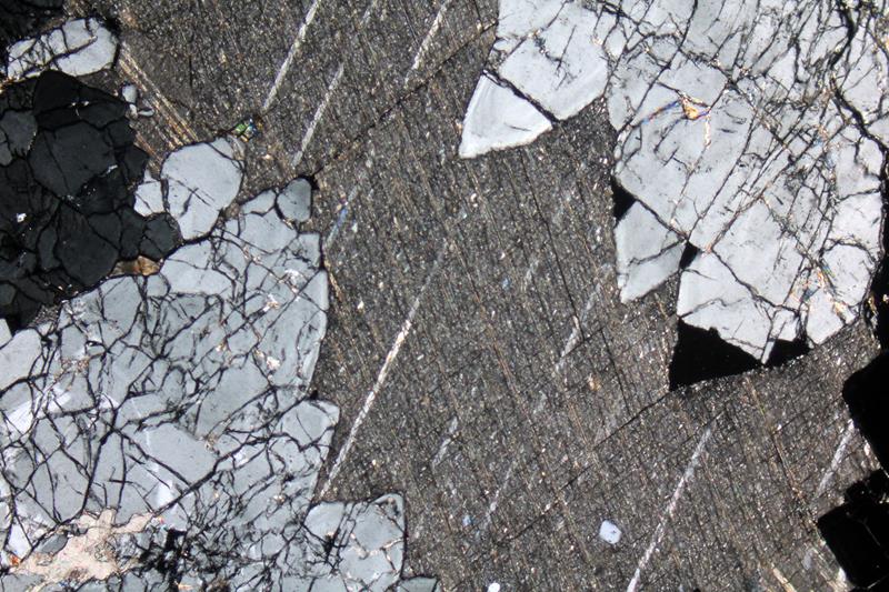
The photo in Figure 16.278 shows a vesuvianite-bearing marble from the Jeffrey Mine in Val-des-Sources, Quebec. The mine is an open-pit asbestos mine that ceased operations around 2001. Vesuvianite, also called idocrase, is one of a number of Ca-Mg silicate minerals that form in calcsilicate rocks. It typically has the green color seen in the photo above. Crystals are tetragonal and a hint of this is visible in the hand specimen.
The plane-polar and crossed-polar views show a different vesuvianite marble. This one is from Gavorrano, Italy. The vesuvianite is well-fractured and has very high relief compared with the calcite. Twins are apparent in both the plane-polar and crossed-polar views of calcite. Vesuvianite in this thin section shows up to 1st-order gray interference colors, but vesuvianite may display lower-order and sometimes anomalous blue interference colors.
The photos below show two other thin sections containing vesuvianite. The rock seen in Figures 16.281 and 16.282 contains high-relief vesuvianite and two dark-green (nearly black) grains of spinel. These minerals appear to be floating in calcite in the XP view. In crossed polars, the vesuvianite shows zoning and up to 1st-order gray interference colors, the spinel is isotropic, and the calcite shows high-order interference colors and twinning.
|
|
|
|
|
|
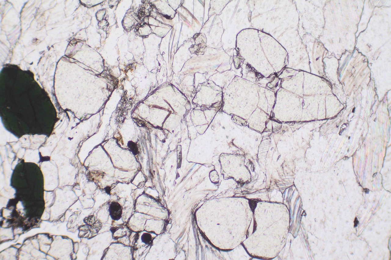
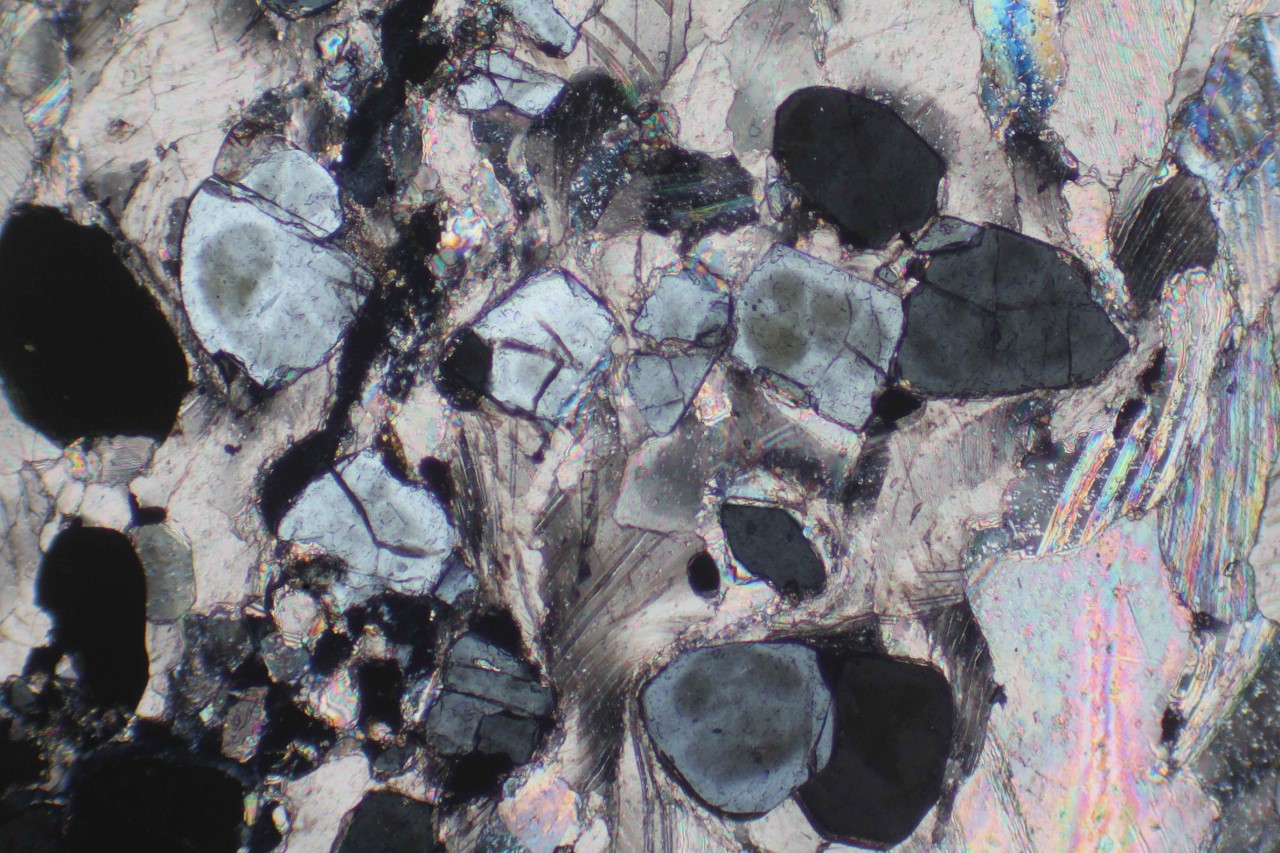
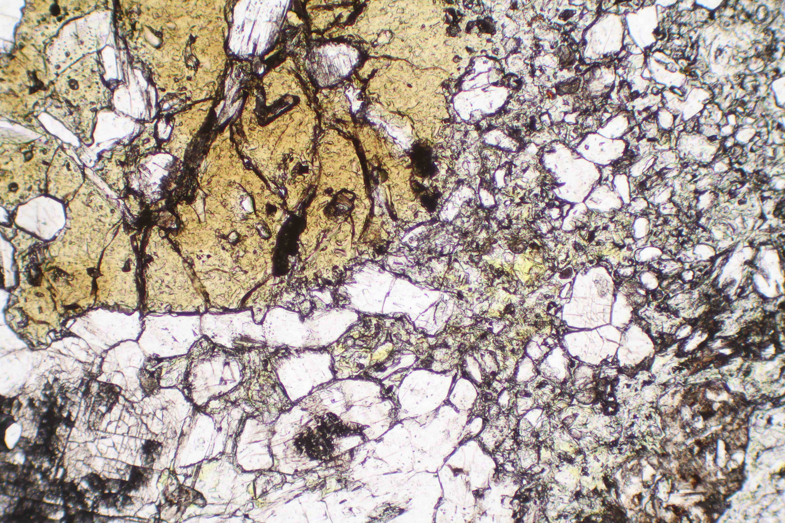

Figures 16.283 and 16.284 show a rock that contains andradite (garnet), vesuvianite, and wollastonite. The andradite is light brown in the PP view and isotropic in crossed polars. The vesuvianite is grayish to light green, and somewhat cloudy, in the PP view. It shows very-low 1st order interference colors in the XP view. If you enlarge the photo, you can see some anomalous blue interference color in some vesuvianite grains. Wollastonite in this rock is clear in PP light and some grains show pyroxene cleavage (near 90o), especially one large grain near the lower left corner. In the XP view, the wollastonite shows up to 1st-order yellow interference colors.
▪Monticellite Marble
The monticellite-bearing marble in Figure 16.285 comes from a quarry in Grand Forks, British Columbia. Besides brown monticellite, white dolomite, and white calcite, this metadolomite contains minor dark-colored diopside. A few small grains of diopside can be seen near the lower-left edge of the specimen.
|
|
|
|
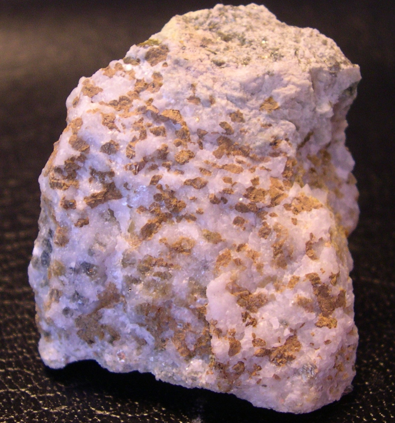

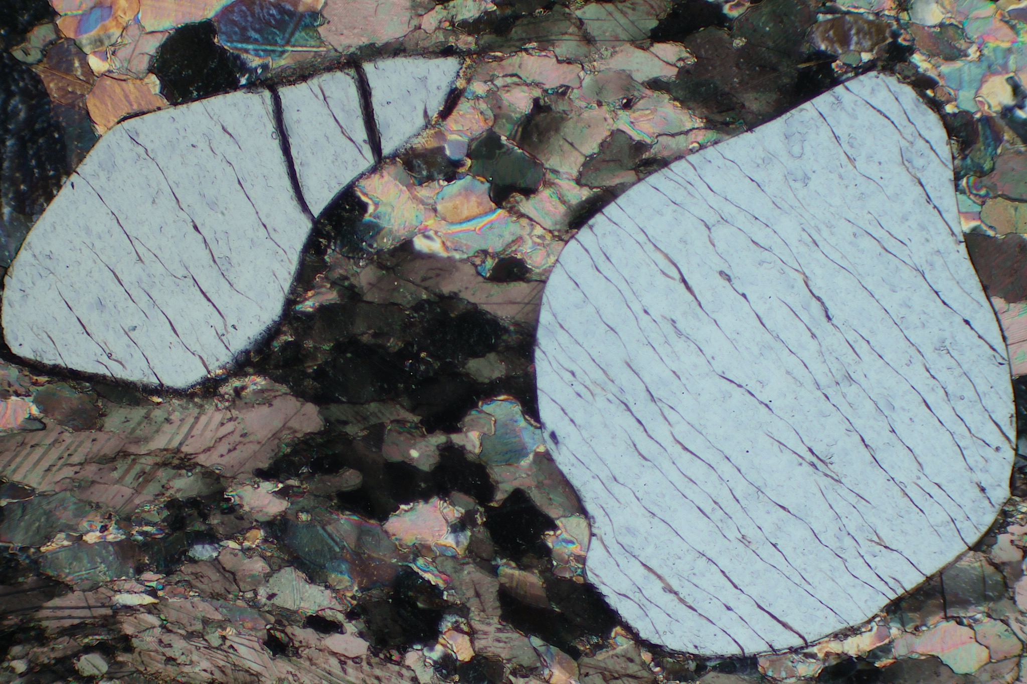
|
|
|
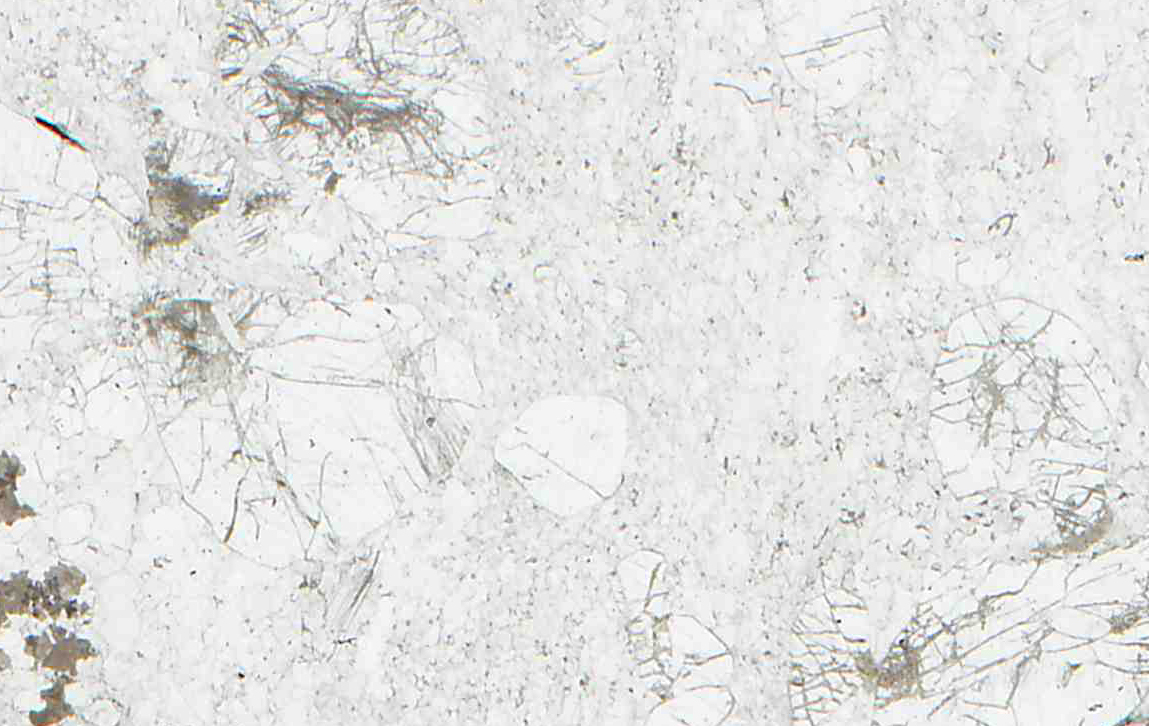
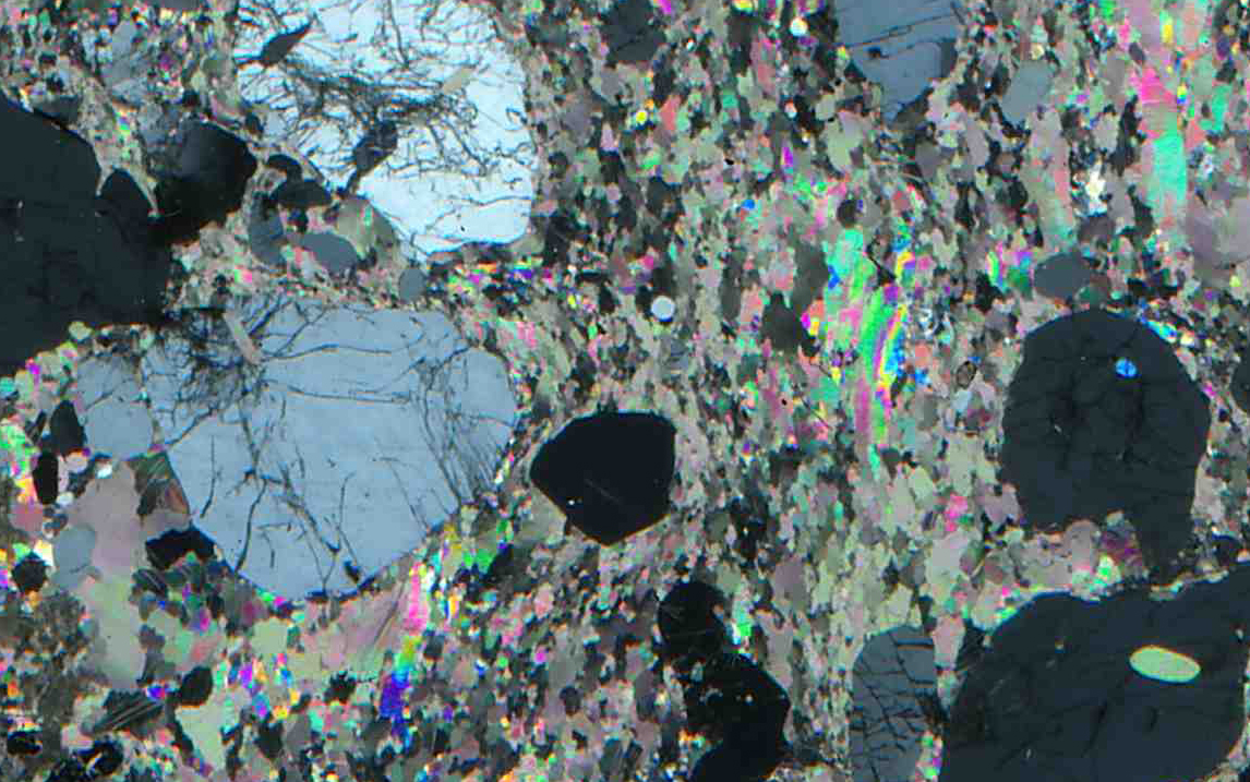
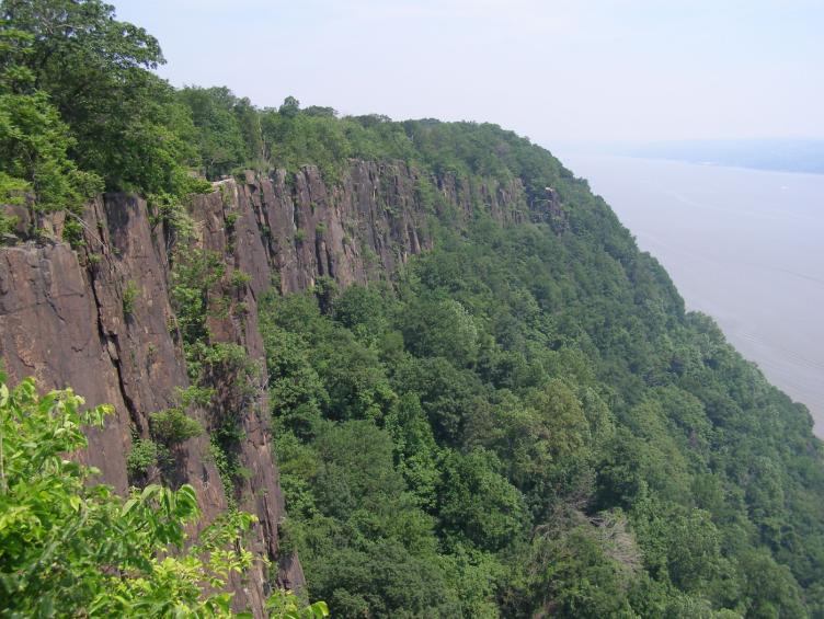
The photos in these figures (16-286 to 16.289) are of the same thin section, but the latter two are six times wider than the first two.
The PP view above (Figure 16.286) shows two large high-relief grains of monticellite surrounded by dolomite and calcite. Monticellite is a member of the olivine group, and like other olivine minerals has no cleavage. The two large grains, however, contain subparallel fractures. In crossed polars, the monticellite shows 1st-order gray interference colors; the surrounding carbonate minerals display high-order pastels. This thin section is a bit thin – normal interference colors for monticellite range up to 1st-order yellow or orange. The photos in Figures 16.287 and 16.288 are of a different part of the same thin section.
● Figure CreditsThe graphics in this chapter were drafted specifically for this book. Many of the photos came from University of North Dakota collections that can be found at: Many other photos came from a number of sources, the most important of which were: The list below gives credits for all photos that did not originate at the University of North Dakota. URLs are listed above for many of these: |


Contents
- Introduction
- 16.1 Plutonic Rocks
- 16.2 Volcanic Rocks
- 16.3 Metamorphic Rocks